Amoeba under microscope 40x
Amoeba Under Microscope 40x. Other important information includes. She needs to calculate the magnification of her drawing. Amoeba under the microscope amoeba is a unicellular organism in the kingdom protozoa. It is a eukaryote and thus has membrane bound cell organelles and protein bound genetic material with a nuclear membrane.
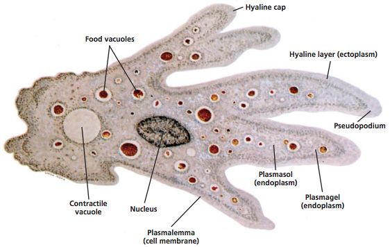 Introduction To Protista Amoeba Carolina Com From carolina.com
Generally the term is used to describe single celled organisms that move in a primitive crawling manner by using temporary false feet known as pseudopods. An amoeba s cell s organelles and cytoplasm are enclosed by the membrane. Ameoba have one pseudopod used for navigation and movement. A tiny blob of colorless jelly with a dark speck inside it this is what an amoeba looks like when seen through a microscope. Cheek cells 40x paramecium 40x amoeba 40x onion skin cells 40x paramecium 40x. Other important information includes.
She needs to calculate the magnification of her drawing.
Food enveloped by the amoeba is stored and digested in vacuoles. A tiny blob of colorless jelly with a dark speck inside it this is what an amoeba looks like when seen through a microscope. It is a eukaryote and thus has membrane bound cell organelles and protein bound genetic material with a nuclear membrane. Amoebas are usually considered among the lowest and most primitive forms of life. This is a great view of cytoplasmic streaming showing how an amoeba moves around by getting its cytoplasm to move to different parts of the cell elongating. Amoeba under the microscope amoeba is a unicellular organism in the kingdom protozoa.
 Source: microbenotes.com
Each amoeba has one or more nuclei and a simple contractile vacuole to maintain osmotic equilibrium. Generally the term is used to describe single celled organisms that move in a primitive crawling manner by using temporary false feet known as pseudopods. Amoeba under the microscope amoeba is a unicellular organism in the kingdom protozoa. Amoebas are usually considered among the lowest and most primitive forms of life. A tiny blob of colorless jelly with a dark speck inside it this is what an amoeba looks like when seen through a microscope.
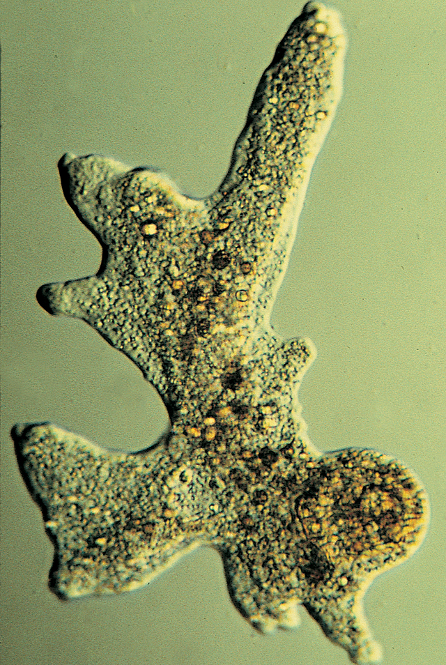 Source: flinnsci.com
Generally the term is used to describe single celled organisms that move in a primitive crawling manner by using temporary false feet known as pseudopods. Worksheet 1 calculations related to the microscope anne viewed an amoeba under the high power 40x objective lens on her microscope. Amoebas are usually considered among the lowest and most primitive forms of life. Generally the term is used to describe single celled organisms that move in a primitive crawling manner by using temporary false feet known as pseudopods. Image of amoeba captured at 400x with a biological.
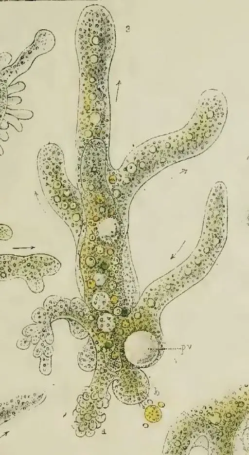 Source: microscopemaster.com
Hd amoeba at 40x 100x 200x and 400x this is a great view of cytoplasmic streaming showing how an amoeba moves around by getting its cytoplasm to move to different parts of the cell elongating. This is a great view of cytoplasmic streaming showing how an amoeba moves around by getting its cytoplasm to move to different parts of the cell elongating. Amoeba moves with their pseudopodia which are a specialized form of the plasma membrane that results in a crawling motion of the organism. Cheek cells 40x paramecium 40x amoeba 40x onion skin cells 40x paramecium 40x. An amoeba s cell s organelles and cytoplasm are enclosed by the membrane.
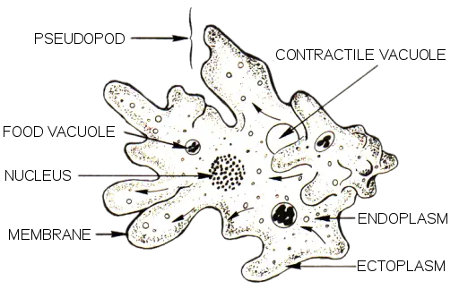 Source: microscopemaster.com
Food enveloped by the amoeba is stored and digested in vacuoles. The colorless jelly is cytoplasm and the dark speck is the nucleus. Each amoeba has one or more nuclei and a simple contractile vacuole to maintain osmotic equilibrium. Amoeba under the microscope amoeba is a unicellular organism in the kingdom protozoa. Ameoba have one pseudopod used for navigation and movement.
 Source: m.youtube.com
Each amoeba has one or more nuclei and a simple contractile vacuole to maintain osmotic equilibrium. Amoeba moves with their pseudopodia which are a specialized form of the plasma membrane that results in a crawling motion of the organism. She drew the following picture of that amoeba. A tiny blob of colorless jelly with a dark speck inside it this is what an amoeba looks like when seen through a microscope. Hd amoeba at 40x 100x 200x and 400x this is a great view of cytoplasmic streaming showing how an amoeba moves around by getting its cytoplasm to move to different parts of the cell elongating.
 Source: youtube.com
She needs to calculate the magnification of her drawing. An amoeba s cell s organelles and cytoplasm are enclosed by the membrane. She drew the following picture of that amoeba. Hd amoeba at 40x 100x 200x and 400x this is a great view of cytoplasmic streaming showing how an amoeba moves around by getting its cytoplasm to move to different parts of the cell elongating. Amoeba under the microscope fixing staining techniques and structure amoeba plural amoebas amoebae is a genus that belongs to kingdom protozoa.
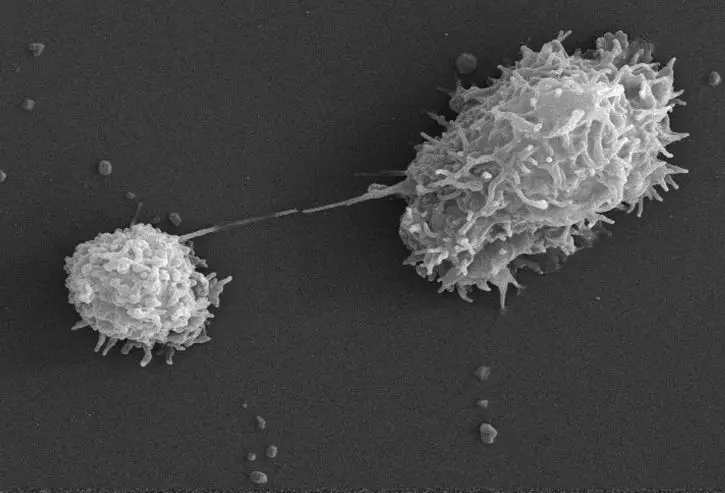 Source: microscopemaster.com
The colorless jelly is cytoplasm and the dark speck is the nucleus. Food enveloped by the amoeba is stored and digested in vacuoles. Other important information includes. Ameoba have one pseudopod used for navigation and movement. A tiny blob of colorless jelly with a dark speck inside it this is what an amoeba looks like when seen through a microscope.
 Source: britannica.com
She drew the following picture of that amoeba. She drew the following picture of that amoeba. She needs to calculate the magnification of her drawing. It is a eukaryote and thus has membrane bound cell organelles and protein bound genetic material with a nuclear membrane. Image of amoeba captured at 400x with a biological.
 Source: carolina.com
Amoeba moves with their pseudopodia which are a specialized form of the plasma membrane that results in a crawling motion of the organism. Amoeba moves with their pseudopodia which are a specialized form of the plasma membrane that results in a crawling motion of the organism. Food enveloped by the amoeba is stored and digested in vacuoles. Cheek cells 40x paramecium 40x amoeba 40x onion skin cells 40x paramecium 40x. This is a great view of cytoplasmic streaming showing how an amoeba moves around by getting its cytoplasm to move to different parts of the cell elongating.
 Source: youtube.com
Worksheet 1 calculations related to the microscope anne viewed an amoeba under the high power 40x objective lens on her microscope. The colorless jelly is cytoplasm and the dark speck is the nucleus. Cheek cells 40x paramecium 40x amoeba 40x onion skin cells 40x paramecium 40x. A tiny blob of colorless jelly with a dark speck inside it this is what an amoeba looks like when seen through a microscope. Ameoba have one pseudopod used for navigation and movement.
 Source: youtube.com
Other important information includes. Amoeba under the microscope amoeba is a unicellular organism in the kingdom protozoa. Cheek cells 40x paramecium 40x amoeba 40x onion skin cells 40x paramecium 40x. This is a great view of cytoplasmic streaming showing how an amoeba moves around by getting its cytoplasm to move to different parts of the cell elongating. A tiny blob of colorless jelly with a dark speck inside it this is what an amoeba looks like when seen through a microscope.

Image of amoeba captured at 400x with a biological. Cheek cells 40x paramecium 40x amoeba 40x onion skin cells 40x paramecium 40x. Other important information includes. Hd amoeba at 40x 100x 200x and 400x this is a great view of cytoplasmic streaming showing how an amoeba moves around by getting its cytoplasm to move to different parts of the cell elongating. She needs to calculate the magnification of her drawing.
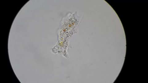 Source: shutterstock.com
Image of amoeba captured at 400x with a biological. Image of amoeba captured at 400x with a biological. She drew the following picture of that amoeba. Hd amoeba at 40x 100x 200x and 400x this is a great view of cytoplasmic streaming showing how an amoeba moves around by getting its cytoplasm to move to different parts of the cell elongating. Ameoba have one pseudopod used for navigation and movement.
 Source: microscope-microscope.org
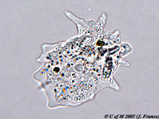
Worksheet 1 calculations related to the microscope anne viewed an amoeba under the high power 40x objective lens on her microscope. Amoebas are usually considered among the lowest and most primitive forms of life. Amoeba moves with their pseudopodia which are a specialized form of the plasma membrane that results in a crawling motion of the organism. This is a great view of cytoplasmic streaming showing how an amoeba moves around by getting its cytoplasm to move to different parts of the cell elongating. The colorless jelly is cytoplasm and the dark speck is the nucleus.
 Source: home.cc.umanitoba.ca
A tiny blob of colorless jelly with a dark speck inside it this is what an amoeba looks like when seen through a microscope. An amoeba s cell s organelles and cytoplasm are enclosed by the membrane. She needs to calculate the magnification of her drawing. A tiny blob of colorless jelly with a dark speck inside it this is what an amoeba looks like when seen through a microscope. Image of amoeba captured at 400x with a biological.
If you find this site adventageous, please support us by sharing this posts to your favorite social media accounts like Facebook, Instagram and so on or you can also bookmark this blog page with the title amoeba under microscope 40x by using Ctrl + D for devices a laptop with a Windows operating system or Command + D for laptops with an Apple operating system. If you use a smartphone, you can also use the drawer menu of the browser you are using. Whether it’s a Windows, Mac, iOS or Android operating system, you will still be able to bookmark this website.

