Bacteria microscope slides
Bacteria Microscope Slides. We also use a variety of stains on bacteria special preparations. Such as meningococcus streptococcus pyogenes and so on. It consists of staphylococcus aureus pus organism sarcina lutea chromogenic rods streptococcus lactis milk souring organism short chains bacillus subtilis hay bacillus smear with bacilli and spores. Here is the procedure.
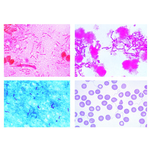 Lieder Pathogenic Bacteria 25 Microscope Slides From optics-pro.com
Such as meningococcus streptococcus pyogenes and so on. Staph infections are caused by a strain of this bacteria. Here is the procedure. Bacteria prepared microscope slides and educational digital images of bacteria including saprophytic bacteria plant pathogens and animal pathogens. In order to view individual bacteria through a light microscope a bacterial smear must be attached to a slide and then stained. Our prepared slides types contain many kinds of bacteria.
In order to view individual bacteria through a light microscope a bacterial smear must be attached to a slide and then stained.
Here is the procedure. Such as meningococcus streptococcus pyogenes and so on. We also use a variety of stains on bacteria special preparations. Our prepared slides types contain many kinds of bacteria. Staph infections are caused by a strain of this bacteria. Bacteria prepared microscope slides and educational digital images of bacteria including saprophytic bacteria plant pathogens and animal pathogens.
 Source: m.carolina.com
You can check your need for our catalog about bacteria. Here presenting microbiology slides of bacteria. First use a wax pencil to draw a circle on the microscope slide to separate each type of bacteria that is going to be sampled. This gram positive bacteria is typically harmless and resides on the skin and in mucous membranes. We also use a variety of stains on bacteria special preparations.
 Source: amazon.com
Staph infections are caused by a strain of this bacteria. It will really beneficial for you. This gram positive bacteria is typically harmless and resides on the skin and in mucous membranes. Bacteria prepared microscope slides and educational digital images of bacteria including saprophytic bacteria plant pathogens and animal pathogens. We also use a variety of stains on bacteria special preparations.
 Source: uk.vwr.com
Staphylococcus staphylococcus microscope prepared slide image at left was captured at 400x magnification. In order to view individual bacteria through a light microscope a bacterial smear must be attached to a slide and then stained. Bacteria prepared microscope slides and educational digital images of bacteria including saprophytic bacteria plant pathogens and animal pathogens. It consists of staphylococcus aureus pus organism sarcina lutea chromogenic rods streptococcus lactis milk souring organism short chains bacillus subtilis hay bacillus smear with bacilli and spores. Our prepared slides types contain many kinds of bacteria.
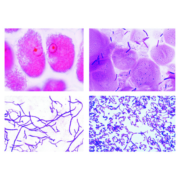 Source: optics-pro.com
Staphylococcus staphylococcus microscope prepared slide image at left was captured at 400x magnification. It consists of staphylococcus aureus pus organism sarcina lutea chromogenic rods streptococcus lactis milk souring organism short chains bacillus subtilis hay bacillus smear with bacilli and spores. It will really beneficial for you. First use a wax pencil to draw a circle on the microscope slide to separate each type of bacteria that is going to be sampled. Such as meningococcus streptococcus pyogenes and so on.
 Source: carolina.com
You can check your need for our catalog about bacteria. We also use a variety of stains on bacteria special preparations. It consists of staphylococcus aureus pus organism sarcina lutea chromogenic rods streptococcus lactis milk souring organism short chains bacillus subtilis hay bacillus smear with bacilli and spores. It will really beneficial for you. Staph infections are caused by a strain of this bacteria.
Source: enasco.com
Here is the procedure. In order to view individual bacteria through a light microscope a bacterial smear must be attached to a slide and then stained. We also use a variety of stains on bacteria special preparations. It consists of staphylococcus aureus pus organism sarcina lutea chromogenic rods streptococcus lactis milk souring organism short chains bacillus subtilis hay bacillus smear with bacilli and spores. Staphylococcus staphylococcus microscope prepared slide image at left was captured at 400x magnification.
 Source: optics-pro.com
First use a wax pencil to draw a circle on the microscope slide to separate each type of bacteria that is going to be sampled. You can check your need for our catalog about bacteria. It will really beneficial for you. Staph infections are caused by a strain of this bacteria. Here presenting microbiology slides of bacteria.
 Source: shopanatomical.com
Such as meningococcus streptococcus pyogenes and so on. We also use a variety of stains on bacteria special preparations. Staphylococcus staphylococcus microscope prepared slide image at left was captured at 400x magnification. Staph infections are caused by a strain of this bacteria. Here presenting microbiology slides of bacteria.
 Source: fishersci.com
We also use a variety of stains on bacteria special preparations. Bacteria prepared microscope slides and educational digital images of bacteria including saprophytic bacteria plant pathogens and animal pathogens. This gram positive bacteria is typically harmless and resides on the skin and in mucous membranes. Staph infections are caused by a strain of this bacteria. Here presenting microbiology slides of bacteria.
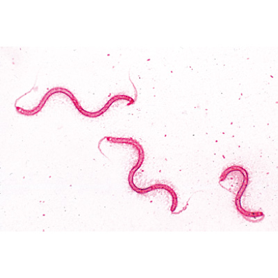 Source: 3bscientific.com
We also use a variety of stains on bacteria special preparations. Staphylococcus staphylococcus microscope prepared slide image at left was captured at 400x magnification. First use a wax pencil to draw a circle on the microscope slide to separate each type of bacteria that is going to be sampled. This gram positive bacteria is typically harmless and resides on the skin and in mucous membranes. Our prepared slides types contain many kinds of bacteria.
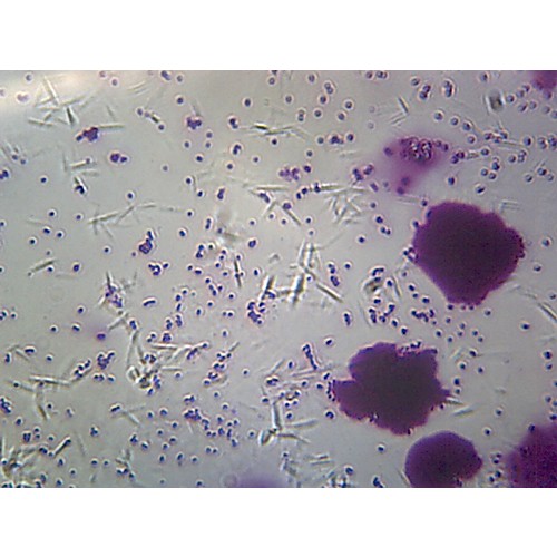 Source: thomassci.com
The sarcina prepared microscope slide at left was captured at 400x magnification. In order to view individual bacteria through a light microscope a bacterial smear must be attached to a slide and then stained. Our prepared slides types contain many kinds of bacteria. We also use a variety of stains on bacteria special preparations. It consists of staphylococcus aureus pus organism sarcina lutea chromogenic rods streptococcus lactis milk souring organism short chains bacillus subtilis hay bacillus smear with bacilli and spores.
Source: store.schoolspecialty.com
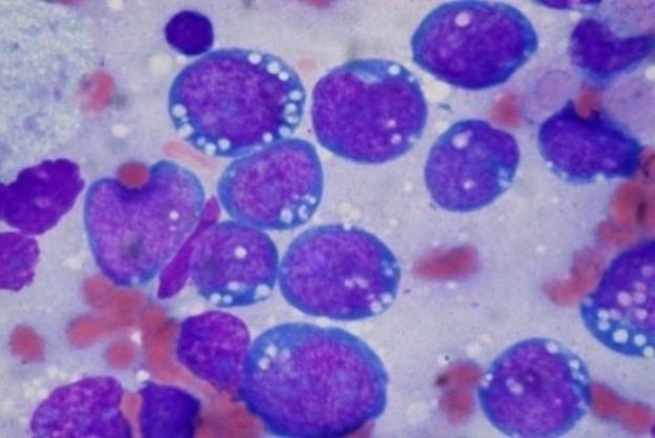
Bacteria prepared microscope slides and educational digital images of bacteria including saprophytic bacteria plant pathogens and animal pathogens. Here presenting microbiology slides of bacteria. This gram positive bacteria is typically harmless and resides on the skin and in mucous membranes. Such as meningococcus streptococcus pyogenes and so on. It consists of staphylococcus aureus pus organism sarcina lutea chromogenic rods streptococcus lactis milk souring organism short chains bacillus subtilis hay bacillus smear with bacilli and spores.
 Source: magnacol.co.uk
Here is the procedure. This gram positive bacteria is typically harmless and resides on the skin and in mucous membranes. It will really beneficial for you. Our prepared slides types contain many kinds of bacteria. The sarcina prepared microscope slide at left was captured at 400x magnification.
 Source: amazon.com
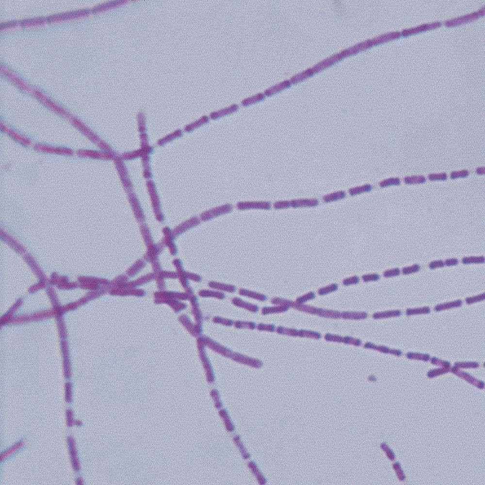
Here is the procedure. You can check your need for our catalog about bacteria. First use a wax pencil to draw a circle on the microscope slide to separate each type of bacteria that is going to be sampled. Such as meningococcus streptococcus pyogenes and so on. This gram positive bacteria is typically harmless and resides on the skin and in mucous membranes.
 Source: carolina.com
Such as meningococcus streptococcus pyogenes and so on. In order to view individual bacteria through a light microscope a bacterial smear must be attached to a slide and then stained. We also use a variety of stains on bacteria special preparations. Staphylococcus staphylococcus microscope prepared slide image at left was captured at 400x magnification. Here is the procedure.
If you find this site adventageous, please support us by sharing this posts to your own social media accounts like Facebook, Instagram and so on or you can also bookmark this blog page with the title bacteria microscope slides by using Ctrl + D for devices a laptop with a Windows operating system or Command + D for laptops with an Apple operating system. If you use a smartphone, you can also use the drawer menu of the browser you are using. Whether it’s a Windows, Mac, iOS or Android operating system, you will still be able to bookmark this website.


