Cardiac muscle slide
Cardiac Muscle Slide. Cardiac muscle is striated involuntary muscle found in the heart wall. Desmosomes hold cells together and gap junctions allow action potentials. Doctor larsencleveland chiropractic college histology anatomy 514laboratory four connective tissue continuedslide 35 cardiac muscle. Cardiac muscle cells are striated meaning they will only contract along their long axis.
 Cardiac Muscle From eugraph.com
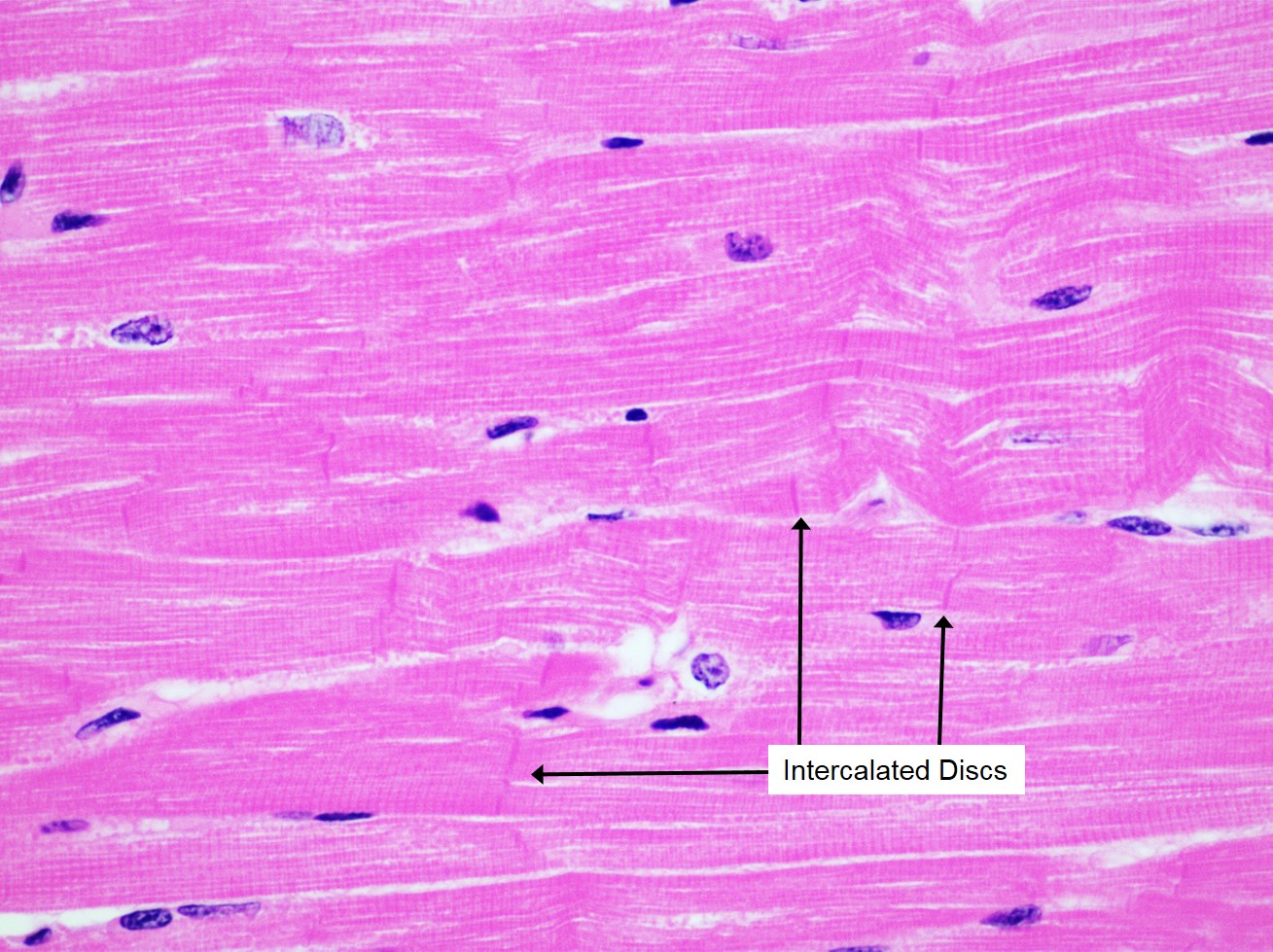
Connect to one another at intercalated discs. Intercalated disks specialized cell cell contacts. Slide 2 cardiac muscle and heart function cardiac muscle fibers are striated sarcomere is the functional unit fibers are branched. Orientation of cardiac muscle fibres. Desmosomes hold cells together and gap junctions allow action potentials. Mohammed abdul hannan hazari assistant professor department of physiology deccan college of medical sciences hyderabad.
3 no transcript 4 cardiac myocyte action potential 5 refractory period.
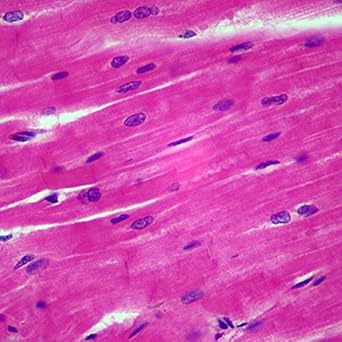
Cardiac muscle cells or cardiomyocytes contain the same contractile filaments as in skeletal muscle. Cardiac muscle is striated involuntary muscle found in the heart wall. 3 no transcript 4 cardiac myocyte action potential 5 refractory period. Now customize the name of a clipboard to store your clips. A cardiac muscle cell typically has one nucleus located near the center. Intercalated disks specialized cell cell contacts.
 Source: medcell.med.yale.edu
Slide 2 cardiac muscle and heart function cardiac muscle fibers are striated sarcomere is the functional unit fibers are branched. The discs contain several gap junctions nuclei are centrally located abundant mitochondria sr is less abundant than in skeletal muscle but greater in density than. Doctor larsencleveland chiropractic college histology anatomy 514laboratory four connective tissue continuedslide 35 cardiac muscle. Contains actin and myosin myofilaments. Cardiac muscle cells have rounded cross sections less than 25 µm in diameter with a centrally located nucleus.
 Source: lab.anhb.uwa.edu.au
Connect to one another at intercalated discs. Clipping is a handy way to collect important slides you want to go back to later. Elongated branching cells containing 1 2 centrally located nuclei. Cardiac muscle cells are cylindrical cells whose ends branch and form junctions with other cardiac muscle cells. 057 ventricle h e webscope 098 1 heart ventricle h e webscope 098n right wall masson webscope 305 heart ventricle h e webscope note.
 Source: education.med.nyu.edu
You just clipped your first slide. Cardiac muscle hd 4 22. Elongated branching cells containing 1 2 centrally located nuclei. In comparison with skeletal muscle note the following differences. Cardiac muscle is striated involuntary muscle found in the heart wall.
 Source: ohiostate.pressbooks.pub
Longitudinal section cardiac muscle cells are smaller in size than skeletal muscle 50 to 250 µm in length. In order to get contraction in two axis the fires wrap around. Cardiac muscle cells or cardiomyocytes contain the same contractile filaments as in skeletal muscle. Clipping is a handy way to collect important slides you want to go back to later. Connect to one another at intercalated discs.
 Source: amazon.com
Cardiac muscle hd 4 22. Connect to one another at intercalated discs. Cardiac muscle is striated involuntary muscle found in the heart wall. Orientation of cardiac muscle fibres. In comparison with skeletal muscle note the following differences.
 Source: pinterest.com
Orientation of cardiac muscle fibres. You just clipped your first slide. 3 no transcript 4 cardiac myocyte action potential 5 refractory period. Intercalated disks specialized cell cell contacts. Now customize the name of a clipboard to store your clips.
 Source: kenhub.com
Doctor larsencleveland chiropractic college histology anatomy 514laboratory four connective tissue continuedslide 35 cardiac muscle. This slide not in glass slide collection cardiac muscle will be studied in the wall of the ventricle of the heart. Mohammed abdul hannan hazari assistant professor department of physiology deccan college of medical sciences hyderabad. Cardiac muscle is striated involuntary muscle found in the heart wall. Cardiac muscle cells have rounded cross sections less than 25 µm in diameter with a centrally located nucleus.
 Source: pinterest.com
Unlike skeletal muscles cardiac muscles have to contract in more than one direction. Orientation of cardiac muscle fibres. A cardiac muscle cell typically has one nucleus located near the center. Now customize the name of a clipboard to store your clips. Cardiac muscle cells or cardiomyocytes contain the same contractile filaments as in skeletal muscle.
 Source: youtube.com
Cardiac muscle is striated involuntary muscle found in the heart wall. Longitudinal section cardiac muscle cells are smaller in size than skeletal muscle 50 to 250 µm in length. Cardiac muscle cells have rounded cross sections less than 25 µm in diameter with a centrally located nucleus. You just clipped your first slide. This slide not in glass slide collection cardiac muscle will be studied in the wall of the ventricle of the heart.
 Source: eugraph.com
Orientation of cardiac muscle fibres. Clipping is a handy way to collect important slides you want to go back to later. Elongated branching cells containing 1 2 centrally located nuclei. Cardiac muscle hd 4 22. Contains actin and myosin myofilaments.
 Source: indiamart.com
057 ventricle h e webscope 098 1 heart ventricle h e webscope 098n right wall masson webscope 305 heart ventricle h e webscope note. Cardiac muscle cells are cylindrical cells whose ends branch and form junctions with other cardiac muscle cells. In comparison with skeletal muscle note the following differences. Now customize the name of a clipboard to store your clips. 3 no transcript 4 cardiac myocyte action potential 5 refractory period.
 Source: stevegallik.org
Unlike skeletal muscles cardiac muscles have to contract in more than one direction. This slide not in glass slide collection cardiac muscle will be studied in the wall of the ventricle of the heart. Contains actin and myosin myofilaments. Elongated branching cells containing 1 2 centrally located nuclei. Clipping is a handy way to collect important slides you want to go back to later.
 Source: histology-world.com
Two cardiac muscle cell nuclei are indicated in the labelled image. Cardiac muscle cells are striated meaning they will only contract along their long axis. 3 no transcript 4 cardiac myocyte action potential 5 refractory period. Now customize the name of a clipboard to store your clips. Mohammed abdul hannan hazari assistant professor department of physiology deccan college of medical sciences hyderabad.
 Source: fishersci.ca
Intercalated disks specialized cell cell contacts. Cardiac muscle cells are cylindrical cells whose ends branch and form junctions with other cardiac muscle cells. Electrically cardiac muscle behaves as single unit. Connect to one another at intercalated discs. You just clipped your first slide.
 Source: amazon.com
Cardiac muscle hd 4 22. In order to get contraction in two axis the fires wrap around. Cardiac muscle cells or cardiomyocytes contain the same contractile filaments as in skeletal muscle. The discs contain several gap junctions nuclei are centrally located abundant mitochondria sr is less abundant than in skeletal muscle but greater in density than. Cardiac muscle cells are striated meaning they will only contract along their long axis.
If you find this site good, please support us by sharing this posts to your favorite social media accounts like Facebook, Instagram and so on or you can also bookmark this blog page with the title cardiac muscle slide by using Ctrl + D for devices a laptop with a Windows operating system or Command + D for laptops with an Apple operating system. If you use a smartphone, you can also use the drawer menu of the browser you are using. Whether it’s a Windows, Mac, iOS or Android operating system, you will still be able to bookmark this website.


