Eye labelled diagram
Eye Labelled Diagram. In biology human biology physics and practical courses in medicine. It is the outer covering a protective tough white layer called the sclera white part of the eye. Glasses contact lenses or surgery can correct the blurry vision it causes. Astigmatism a condition in which the lens is warped causing images not to focus properly on the retina.
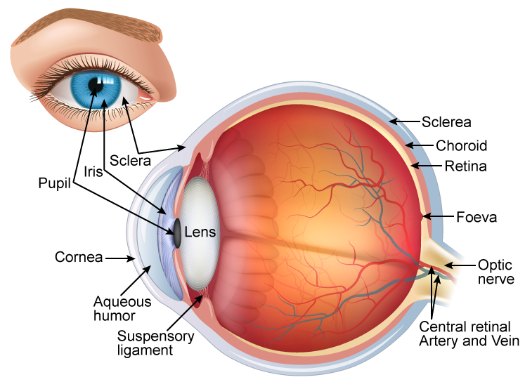 Draw A Neat And Labelled Diagram Of Structure Of The Human Eye Science Topperlearning Com Slwbyx77 From topperlearning.com
Glasses contact lenses or surgery can correct the blurry vision it causes. In addition to the eyeball itself the orbit contains the muscles that move the eye blood vessels and nerves. Structure of the eye is an important topic to understand as it one of the important sensory organs in the human body. The eye is a hollow spherical structure measuring about 2 5 cm in diameter. The human eye is composed of many different parts that work together to interpret the world around us. Aqueous humor the clear watery fluid inside the eye.
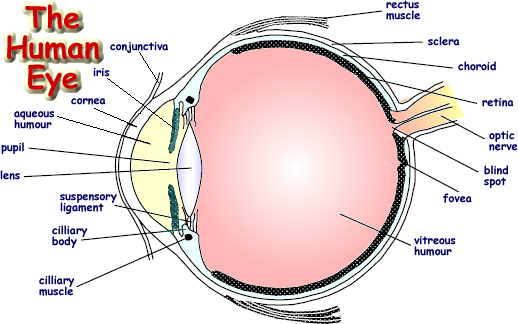
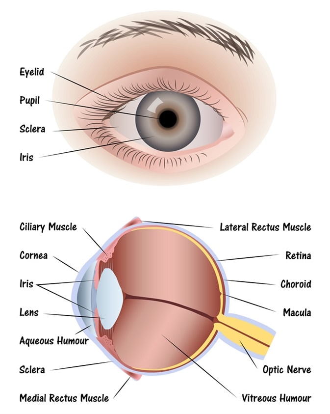
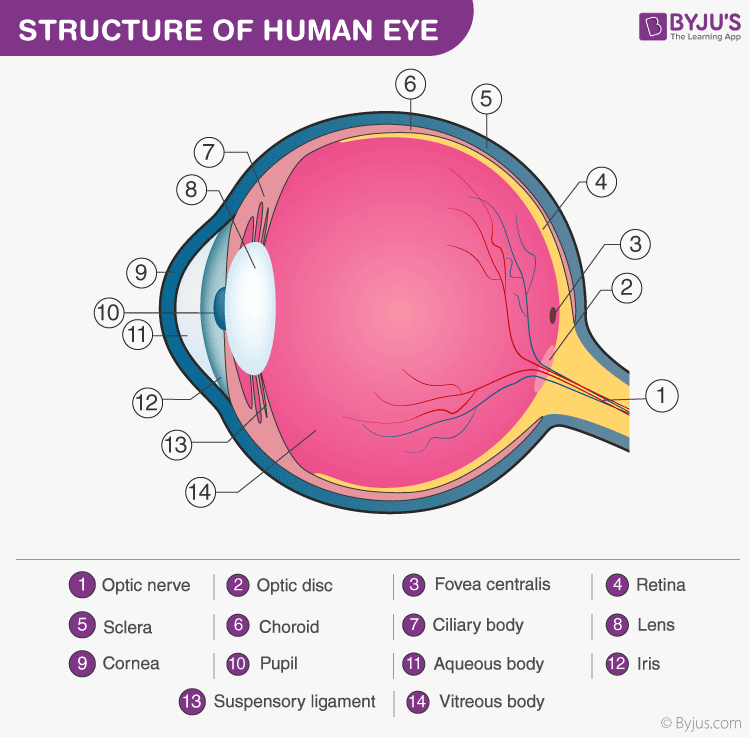
The cornea the pupil the iris the lens the vitreous humor the retina and the sclera.
Structure of human eye. Glasses contact lenses or surgery can correct the blurry vision it causes. The human eye is composed of many different parts that work together to interpret the world around us. It is mainly responsible for vision differentiation of colour the human eye can differentiate approximately 10 12 million colours and maintaining the biological clock of the human body. A closer look at the parts of the eye by liz segre when surveyed about the five senses sight hearing taste smell and touch people consistently report that their eyesight is the mode of perception they value and fear losing most. Astigmatism a condition in which the lens is warped causing images not to focus properly on the retina.
 Source: pinterest.com
Diagram of the choroid iris and ciliary body. Cones cells the in the retina that sense color. Binocular vision the coordinated use of two eyes which gives the ability to see the world in three dimensions 3d. What you want to interpret as a major part of the human eye is somewhat up to the individual but in general there are seven parts of the human eye. The middle vascular coat choroid ciliary body iris.
 Source: topperlearning.com
Glasses contact lenses or surgery can correct the blurry vision it causes. Aqueous humor the clear watery fluid inside the eye. What you want to interpret as a major part of the human eye is somewhat up to the individual but in general there are seven parts of the human eye. The inner sensorineural layer is known as the retina. The eye is a hollow spherical structure measuring about 2 5 cm in diameter.
 Source: diagramgrimj.camperlot.it
Light enters the eye through the cornea. Structure of human eye. In this article we will discuss about the structure of human eye. Binocular vision the coordinated use of two eyes which gives the ability to see the world in three dimensions 3d. It consists of the following parts.

The eye is cushioned within the orbit by pads of fat. Let s take a closer look at each of these. Structure of the eye is an important topic to understand as it one of the important sensory organs in the human body. The inner sensorineural layer is known as the retina. The eye is cushioned within the orbit by pads of fat.
 Source: boozmanhof.com
The front transparent part of the sclera is called cornea. It provides nutrients to the eye. The iris is the central diaphragm at the front of the eye. It can open or close to widen or narrow the central aperture known as the pupil. A closer look at the parts of the eye by liz segre when surveyed about the five senses sight hearing taste smell and touch people consistently report that their eyesight is the mode of perception they value and fear losing most.
 Source: rnib.org.uk
Light enters the eye through the cornea. It can open or close to widen or narrow the central aperture known as the pupil. It is the outer covering a protective tough white layer called the sclera white part of the eye. The middle vascular coat choroid ciliary body iris. Let s take a closer look at each of these.
 Source: retina-international.org
The eye is a hollow spherical structure measuring about 2 5 cm in diameter. The outer fibrous coat sclera cornea. It can open or close to widen or narrow the central aperture known as the pupil. The cornea the pupil the iris the lens the vitreous humor the retina and the sclera. A closer look at the parts of the eye by liz segre when surveyed about the five senses sight hearing taste smell and touch people consistently report that their eyesight is the mode of perception they value and fear losing most.
 Source: news-medical.net
The eye is a hollow spherical structure measuring about 2 5 cm in diameter. It can open or close to widen or narrow the central aperture known as the pupil. Astigmatism a condition in which the lens is warped causing images not to focus properly on the retina. The inner sensorineural layer is known as the retina. Causes loss of central vision as you get older.
 Source: thoughtco.com
It is mainly responsible for vision differentiation of colour the human eye can differentiate approximately 10 12 million colours and maintaining the biological clock of the human body. In this article we will discuss about the structure of human eye. Structure of human eye. It is mainly responsible for vision differentiation of colour the human eye can differentiate approximately 10 12 million colours and maintaining the biological clock of the human body. The middle vascular coat choroid ciliary body iris.
 Source: byjus.com
In addition to the eyeball itself the orbit contains the muscles that move the eye blood vessels and nerves. The middle vascular coat choroid ciliary body iris. The eye is a hollow spherical structure measuring about 2 5 cm in diameter. In addition to the eyeball itself the orbit contains the muscles that move the eye blood vessels and nerves. The human eye is composed of many different parts that work together to interpret the world around us.

The eye is cushioned within the orbit by pads of fat. Often called lazy eye this condition starts in childhood one eye sees better than the. The middle vascular coat choroid ciliary body iris. Glasses contact lenses or surgery can correct the blurry vision it causes. What you want to interpret as a major part of the human eye is somewhat up to the individual but in general there are seven parts of the human eye.
 Source: researchgate.net
Its wall is composed of three coats. The cornea the pupil the iris the lens the vitreous humor the retina and the sclera. The lacrimal gland produces tears that help lubricate and moisten the. The iris is the central diaphragm at the front of the eye. What you want to interpret as a major part of the human eye is somewhat up to the individual but in general there are seven parts of the human eye.
 Source: pinterest.com
In this article we will discuss about the structure of human eye. In addition to the eyeball itself the orbit contains the muscles that move the eye blood vessels and nerves. In this article we will discuss about the structure of human eye. Often called lazy eye this condition starts in childhood one eye sees better than the. Glasses contact lenses or surgery can correct the blurry vision it causes.
 Source: shutterstock.com
It is mainly responsible for vision differentiation of colour the human eye can differentiate approximately 10 12 million colours and maintaining the biological clock of the human body. Glasses contact lenses or surgery can correct the blurry vision it causes. In biology human biology physics and practical courses in medicine. Its wall is composed of three coats. It consists of the following parts.
 Source: emedicinehealth.com
Let s take a closer look at each of these. Structure of human eye. It is mainly responsible for vision differentiation of colour the human eye can differentiate approximately 10 12 million colours and maintaining the biological clock of the human body. Astigmatism a condition in which the lens is warped causing images not to focus properly on the retina. The inner sensorineural layer is known as the retina.
If you find this site convienient, please support us by sharing this posts to your favorite social media accounts like Facebook, Instagram and so on or you can also bookmark this blog page with the title eye labelled diagram by using Ctrl + D for devices a laptop with a Windows operating system or Command + D for laptops with an Apple operating system. If you use a smartphone, you can also use the drawer menu of the browser you are using. Whether it’s a Windows, Mac, iOS or Android operating system, you will still be able to bookmark this website.
