Gram staining diagram
Gram Staining Diagram. The name comes from the danish bacteriologist hans christian gram who developed the technique. Gram positive bacteria and gram negative bacteria. Gram stain or gram staining also called gram s method is a method of staining used to distinguish and classify bacterial species into two large groups. Gram staining procedure is illustrated in fig 5 2.
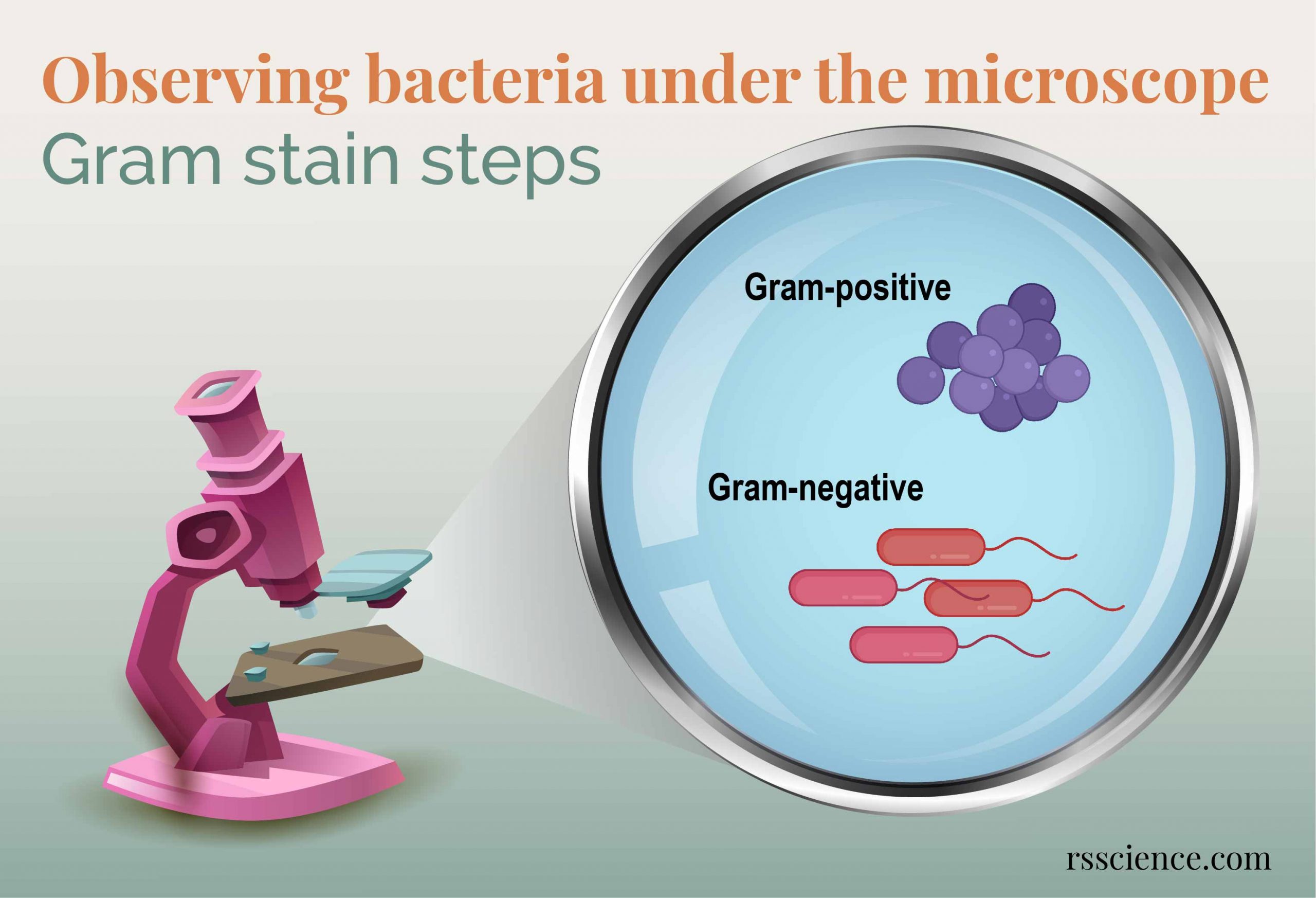 Observing Bacteria Under The Microscope Gram Stain Steps Rs Science From rsscience.com
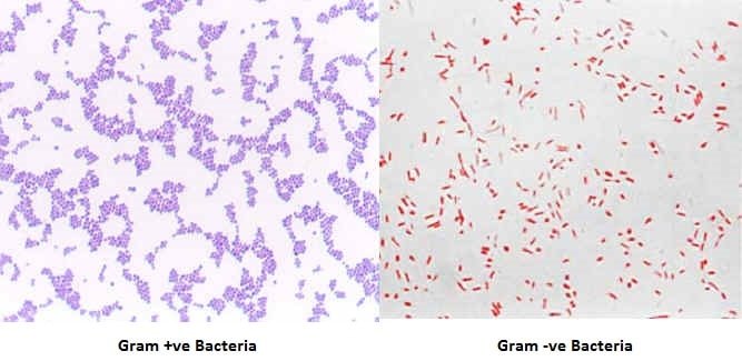
In addition this stain also allows determination of cell morphology size and arrangement. Gram positive bacteria thick layer of peptidoglycan 90 of cell wall stains purple. At lower concentrations the gram stain of a clinical specimen seldom reveals organisms even if the culture is positive. The name comes from the danish bacteriologist hans christian gram who developed the technique. Gram staining procedure is illustrated in fig 5 2. The gram staining technique is the most important and widely used microbiological differential staining technique.
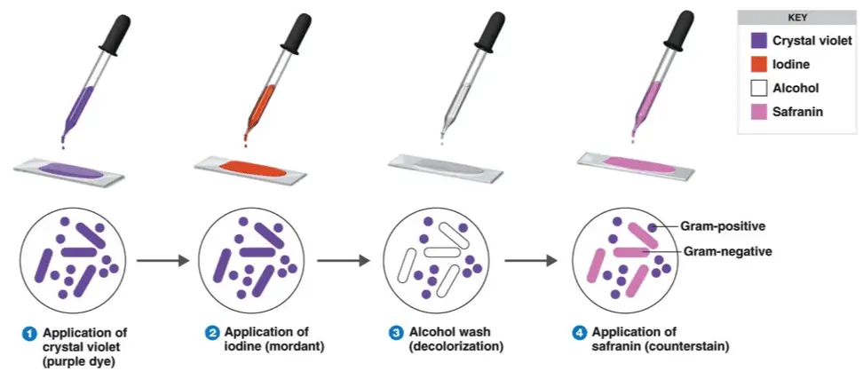
Gram staining procedure is illustrated in fig 5 2.
Gram staining principle. Gram stain or gram staining also called gram s method is a method of staining used to distinguish and classify bacterial species into two large groups. At lower concentrations the gram stain of a clinical specimen seldom reveals organisms even if the culture is positive. To provide information about the gram reaction of the bacteria as well as the cell morphology and cellular arrangement. List the primary dye counter stain and decolorizer used in this stain. Gram positive cells have a thick layer of peptidoglycan in the cell wall that retains the primary st.
 Source: youtube.com
To be visible on a slide organisms that stain by the gram method must be present in concentrations of a minimum of 104to 105organisms ml of unconcentrated staining fluid. Christian gram in 1884 and categorizes bacteria according to their gram character gram positive or gram negative. It is one of the most important and widely used differential staining techniques in microbiology. This differential staining procedure separates most bacteria into two groups on the basis of cell wall composition. Gram stain or gram staining also called gram s method is a method of staining used to distinguish and classify bacterial species into two large groups.
 Source: en.wikipedia.org
Gram staining principle. In addition this stain also allows determination of cell morphology size and arrangement. This test differentiate the bacteria into gram positive and gram negative bacteria which helps in the classification and differentiations of microorganisms. Gram staining principle. Gram staining differentiates bacteria by the chemical and physical properties of their cell walls.
 Source: researchgate.net
It is one of the most important and widely used differential staining techniques in microbiology. Christian gram in 1884 and categorizes bacteria according to their gram character gram positive or gram negative. The gram staining technique is the most important and widely used microbiological differential staining technique. Cell wall structure identifies either cell is gram positive or negative in nature. During the procedure when we stained by primary stain and secure it by a mordant.
 Source: microbiologyinfo.com
To provide information about the gram reaction of the bacteria as well as the cell morphology and cellular arrangement. To provide information about the gram reaction of the bacteria as well as the cell morphology and cellular arrangement. Gram positive cells have a thick layer of peptidoglycan in the cell wall that retains the primary st. Gram stain or gram staining also called gram s method is a method of staining used to distinguish and classify bacterial species into two large groups. In addition this stain also allows determination of cell morphology size and arrangement.
 Source: microbeonline.com
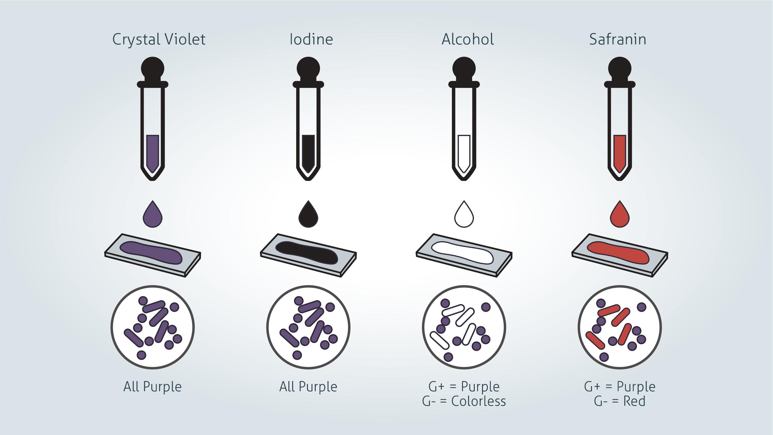
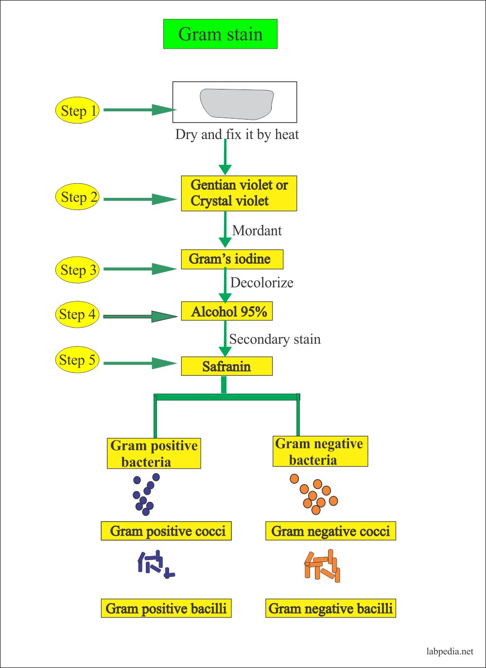
In addition this stain also allows determination of cell morphology size and arrangement. The name comes from the danish bacteriologist hans christian gram who developed the technique. Gram staining procedure is illustrated in fig 5 2. Staining type 3. The primary dye is crystal violet the counterstain is gram safranin and the decolourizer is alcohol acetone.
 Source: laboratoryinfo.com
It is one of the most important and widely used differential staining techniques in microbiology. List the primary dye counter stain and decolorizer used in this stain. Staining type 3. The gram staining technique is the most important and widely used microbiological differential staining technique. The primary dye is crystal violet the counterstain is gram safranin and the decolourizer is alcohol acetone.
 Source: cen.acs.org
It was developed by dr. The primary dye is crystal violet the counterstain is gram safranin and the decolourizer is alcohol acetone. Cell wall structure identifies either cell is gram positive or negative in nature. Gram positive bacteria and gram negative bacteria. Christian gram in 1884 and categorizes bacteria according to their gram character gram positive or gram negative.
 Source: technologynetworks.com
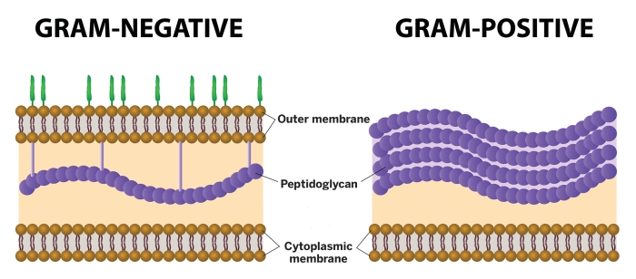
During the procedure when we stained by primary stain and secure it by a mordant. Gram positive bacteria thick layer of peptidoglycan 90 of cell wall stains purple. Gram negative bacteria thin layer of peptidoglycan 10 of cell wall and high lipid content stains red pink. Christian gram in 1884 and categorizes bacteria according to their gram character gram positive or gram negative. Gram positive bacteria and gram negative bacteria.
 Source: researchgate.net
Cell wall structure identifies either cell is gram positive or negative in nature. The gram staining technique is the most important and widely used microbiological differential staining technique. To provide information about the gram reaction of the bacteria as well as the cell morphology and cellular arrangement. Staining type 3. This differential staining procedure separates most bacteria into two groups on the basis of cell wall composition.
 Source: pinterest.com
This technique was introduced in 1884 by danish physician christian gram. Gram stain or gram staining also called gram s method is a method of staining used to distinguish and classify bacterial species into two large groups. To provide information about the gram reaction of the bacteria as well as the cell morphology and cellular arrangement. Cell wall structure identifies either cell is gram positive or negative in nature. This test differentiate the bacteria into gram positive and gram negative bacteria which helps in the classification and differentiations of microorganisms.
 Source: quizlet.com
This test differentiate the bacteria into gram positive and gram negative bacteria which helps in the classification and differentiations of microorganisms. Gram stain or gram staining also called gram s method is a method of staining used to distinguish and classify bacterial species into two large groups. Gram positive cells have a thick layer of peptidoglycan in the cell wall that retains the primary st. The primary dye is crystal violet the counterstain is gram safranin and the decolourizer is alcohol acetone. Gram positive bacteria and gram negative bacteria.
 Source: courses.lumenlearning.com
This test differentiate the bacteria into gram positive and gram negative bacteria which helps in the classification and differentiations of microorganisms. The gram staining technique is the most important and widely used microbiological differential staining technique. Cell wall structure identifies either cell is gram positive or negative in nature. It was developed by dr. At lower concentrations the gram stain of a clinical specimen seldom reveals organisms even if the culture is positive.
 Source: rsscience.com
To provide information about the gram reaction of the bacteria as well as the cell morphology and cellular arrangement. List the primary dye counter stain and decolorizer used in this stain. To be visible on a slide organisms that stain by the gram method must be present in concentrations of a minimum of 104to 105organisms ml of unconcentrated staining fluid. The name comes from the danish bacteriologist hans christian gram who developed the technique. In addition this stain also allows determination of cell morphology size and arrangement.
 Source: labpedia.net
This differential staining procedure separates most bacteria into two groups on the basis of cell wall composition. Gram staining differentiates bacteria by the chemical and physical properties of their cell walls. List the primary dye counter stain and decolorizer used in this stain. At lower concentrations the gram stain of a clinical specimen seldom reveals organisms even if the culture is positive. Gram staining procedure is illustrated in fig 5 2.
 Source: technologynetworks.com
Christian gram in 1884 and categorizes bacteria according to their gram character gram positive or gram negative. This differential staining procedure separates most bacteria into two groups on the basis of cell wall composition. This technique was introduced in 1884 by danish physician christian gram. The name comes from the danish bacteriologist hans christian gram who developed the technique. The primary dye is crystal violet the counterstain is gram safranin and the decolourizer is alcohol acetone.
If you find this site good, please support us by sharing this posts to your favorite social media accounts like Facebook, Instagram and so on or you can also bookmark this blog page with the title gram staining diagram by using Ctrl + D for devices a laptop with a Windows operating system or Command + D for laptops with an Apple operating system. If you use a smartphone, you can also use the drawer menu of the browser you are using. Whether it’s a Windows, Mac, iOS or Android operating system, you will still be able to bookmark this website.


