Human squamous epithelium smear
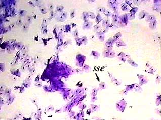
Human Squamous Epithelium Smear. Epithelial tissue covers or lines body surfaces as well as serving to absorb filtrate protect and secrete various substances. These cells make up the outer layer of skin as well as lining the internal organs. Mammal transitional epithelium sec. The lining of the human mouth is a standard example of stratified squamous epithelium.
 Human Stratified Squamous Epithelium Slide Smear Carolina Com From m.carolina.com
Mammal transitional epithelium sec. Human simple ciliated epithelium c s h e microscope slide section of fallopian tube. They are called squamous or scaly for their distinctive thin flat shape. 406 256 0990 or live chat in. Mammal transitional epithelium sec. 7 µm h e microscope slide from cat or dog ureter.
The lining of the human mouth is a standard example of stratified squamous epithelium.
Human stratified squamous epithelium smear microscope slide smear of human cheek cells stained to show general structures of animal cells. Ages 8 in stock ready to ship need it fast. Smear of epithelial cells from inside the mouth. The lining of the human mouth is a standard example of stratified squamous epithelium. See delivery options in cart. Human stratified squamous epithelium slide smear.
 Source: magscope.com
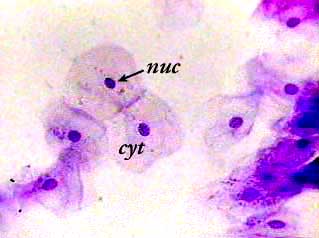
The flat cells shown on the slide form several layers that make. Mammal transitional epithelium sec. Pms5 121 is stained with hematoxylin and eosin. Human stratified squamous epithelium smear microscope slide smear of human cheek cells stained to show general structures of animal cells. The nuclei stain purple and the cytoplasm stains pink.
 Source: magscope.com
Human stratified squamous epithelium smear microscope slide smear of human cheek cells stained to show general structures of animal cells. The nuclei stain purple and the cytoplasm stains pink. 7 µm h e microscope slide from cat or dog ureter. Epithelial tissue covers or lines body surfaces as well as serving to absorb filtrate protect and secrete various substances. They are called squamous or scaly for their distinctive thin flat shape.
 Source: ouhsc.edu
The flat cells shown on the slide form several layers that make up this type of epithelial tissue. The lining of the human mouth is a standard example of stratified squamous epithelium. The lining of the human mouth is a standard example of stratified squamous epithelium. The flat cells shown on the slide form several layers that make up this type of epithelial tissue. They are called squamous or scaly for their distinctive thin flat shape.
 Source: amazon.com
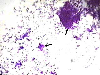
The lining of the human mouth is a standard example of stratified squamous epithelium. Use the isolated cheek cells on this prepared slide to illustrate the morphology of typical squamous cells. Pms5 121 is stained with hematoxylin and eosin. 406 256 0990 or live chat in. In this slide you can see bacteria from the mouth which stains purple as well as the epithelium squamous human mouth cheek cells.
 Source: leermiddelen.be
Human stratified squamous epithelium smear microscope slide smear of human cheek cells stained to show general structures of animal cells. Pms5 121 is stained with hematoxylin and eosin. The nuclei stain purple and the cytoplasm stains pink. Mammal transitional epithelium sec. The lining of the human mouth is a standard example of stratified squamous epithelium.
 Source: austincc.edu
The flat cells shown on the slide form several layers that make up this type of epithelial tissue. 7 µm h e microscope slide from cat or dog ureter. Human squamous epithelium slide smear. 406 256 0990 or live chat in. Mammal transitional epithelium sec.
 Source: homesciencetools.com
Mammal transitional epithelium sec. Smear of epithelial cells from inside the mouth. Pms5 121 is stained with hematoxylin and eosin. Human simple ciliated epithelium c s h e microscope slide section of fallopian tube. Mammal transitional epithelium sec.
 Source: shutterstock.com
Epithelial tissue covers or lines body surfaces as well as serving to absorb filtrate protect and secrete various substances. 7 µm h e microscope slide from cat or dog ureter. The lining of the human mouth is a standard example of stratified squamous epithelium. The nuclei stain purple and the cytoplasm stains pink. Epithelial tissue covers or lines body surfaces as well as serving to absorb filtrate protect and secrete various substances.
 Source: researchgate.net
Human stratified squamous epithelium slide smear. Use the isolated cheek cells on this prepared slide to illustrate the morphology of typical squamous cells. Pms5 121 is stained with hematoxylin and eosin. Epithelial tissue covers or lines body surfaces as well as serving to absorb filtrate protect and secrete various substances. See delivery options in cart.
 Source: dreamstime.com
Human squamous epithelium slide smear. In this slide you can see bacteria from the mouth which stains purple as well as the epithelium squamous human mouth cheek cells. The lining of the human mouth is a standard example of stratified squamous epithelium. 7 µm h e microscope slide from cat or dog ureter. Ages 8 in stock ready to ship need it fast.
 Source: austincc.edu
Human stratified squamous epithelium slide smear. The lining of the human mouth is a standard example of stratified squamous epithelium. Human stratified squamous epithelium smear microscope slide smear of human cheek cells stained to show general structures of animal cells. Smear of epithelial cells from inside the mouth. They are called squamous or scaly for their distinctive thin flat shape.
 Source: m.carolina.com
Mammal transitional epithelium sec. The flat cells shown on the slide form several layers that make. The lining of the human mouth is a standard example of stratified squamous epithelium. The lining of the human mouth is a standard example of stratified squamous epithelium. Human stratified squamous epithelium smear microscope slide smear of human cheek cells stained to show general structures of animal cells.
 Source: medcell.med.yale.edu
Pms5 121 is stained with hematoxylin and eosin. 7 µm h e microscope slide from cat or dog ureter. Human stratified squamous epithelium slide smear. They are called squamous or scaly for their distinctive thin flat shape. These cells make up the outer layer of skin as well as lining the internal organs.
 Source: magscope.com
Epithelial tissue covers or lines body surfaces as well as serving to absorb filtrate protect and secrete various substances. The flat cells shown on the slide form several layers that make up this type of epithelial tissue. 7 µm h e microscope slide from cat or dog ureter. Human simple ciliated epithelium c s h e microscope slide section of fallopian tube. 7 µm h e microscope slide from cat or dog ureter.
 Source: austincc.edu
Mammal transitional epithelium sec. The flat cells shown on the slide form several layers that make. Human stratified squamous epithelium slide smear. Human simple ciliated epithelium c s h e microscope slide section of fallopian tube. 406 256 0990 or live chat in.
If you find this site good, please support us by sharing this posts to your favorite social media accounts like Facebook, Instagram and so on or you can also save this blog page with the title human squamous epithelium smear by using Ctrl + D for devices a laptop with a Windows operating system or Command + D for laptops with an Apple operating system. If you use a smartphone, you can also use the drawer menu of the browser you are using. Whether it’s a Windows, Mac, iOS or Android operating system, you will still be able to bookmark this website.

