Leaf under microscope
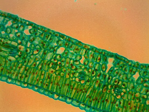
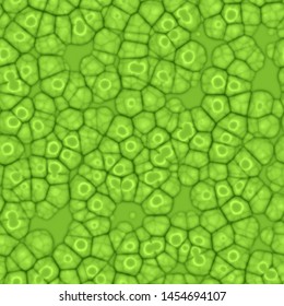
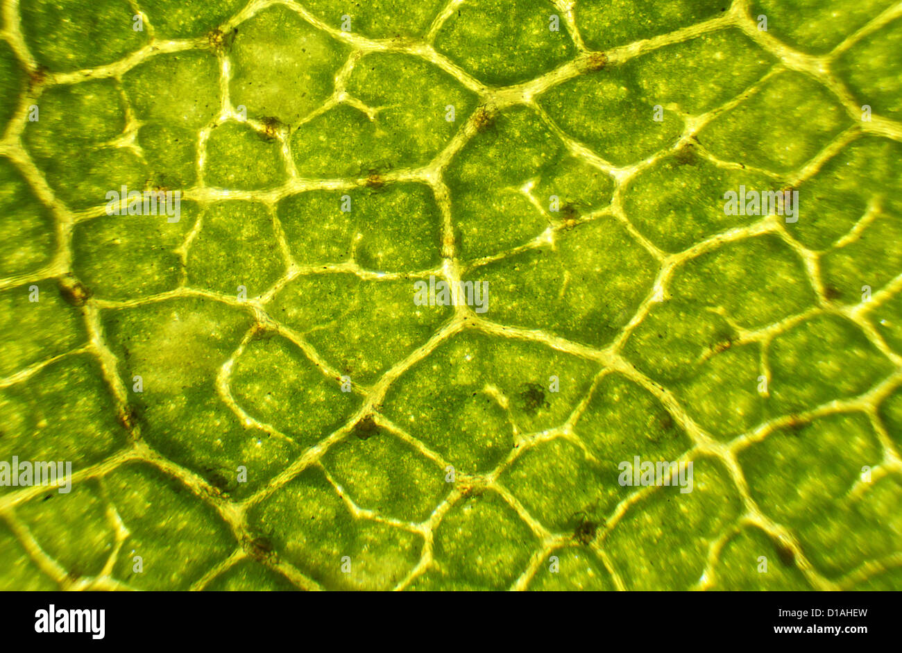
Leaf Under Microscope. Have students paint a thin layer of clear nail polish on the under side of the leaf in a 1 cm x 1 cm square. If you are trying out a new leaf type be sure to test it yourself before doing it with students. Put a drop of water on the microscope slides. Viewing the leaf under the microscope shows different types of cells that serve various functions.
 Leaf Cells Under Microscope Micrograph Leaf Under A Microscope Stock Photo Picture And Royalty Free Image Image 85392618 From 123rf.com
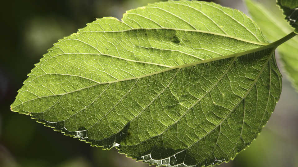
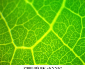
If you are trying out a new leaf type be sure to test it yourself before doing it with students. Put on a coverslip and ready to see. Leaf hairs of a mullein plant verbascum under the microscope. You can see the veins of it under 100x magnification. Using a microscope it s possible to view and identify these cells and how they are arranged epidermal cells spongy cells etc. To do this a compound microscope is required given that it allows for higher magnification.
Do this step next to a drop of water so the spores can be released into water.
You can see the veins of it under 100x magnification. Use tweezers to hold the frond and use a dissecting needle to open sorus. Using a microscope it s possible to view and identify these cells and how they are arranged epidermal cells spongy cells etc. Have students paint a thin layer of clear nail polish on the under side of the leaf in a 1 cm x 1 cm square. Do this step next to a drop of water so the spores can be released into water. Give each student a leaf and a microscope slide.

Viewing the leaf under the microscope shows different types of cells that serve various functions. Dicot leaf cross section dorsiventral leaf anatomical structure of a dicot leaf ixora mangifera hibiscus ø leaves are structurally well adapted to perform the photosynthesis transpiration and gaseous exchange. However any broad flat leaf should work. Leaf hairs of a mullein plant verbascum under the microscope. Also called leaf lamina is the flattened expanded part of the leaf chiefly composed of mesophyll tissue and vascular bundles.
 Source: youtube.com
ø a leaf composed of. Dicot leaf cross section dorsiventral leaf anatomical structure of a dicot leaf ixora mangifera hibiscus ø leaves are structurally well adapted to perform the photosynthesis transpiration and gaseous exchange. Leaf hairs of a mullein plant verbascum under the microscope. Viewing the leaf under the microscope shows different types of cells that serve various functions. Micrograph leaf under a microscope organ producing oxygen and carbon dioxide the process.
 Source: pinterest.com
If you are trying out a new leaf type be sure to test it yourself before doing it with students. Put on a coverslip and ready to see. We put a leaf lemon tree under the microscope. Diagrams of monocot leaf under the microscope author. Diagrams of monocot leaf under the microscope keywords.
 Source: microscopemaster.com
You can see the veins of it under 100x magnification. To do this a compound microscope is required given that it allows for higher magnification. We put a leaf lemon tree under the microscope. Have students paint a thin layer of clear nail polish on the under side of the leaf in a 1 cm x 1 cm square. Diagrams of monocot leaf under the microscope author.
 Source: shutterstock.com
Diagrams of monocot leaf under the microscope author. Micrograph leaf under a microscope organ producing oxygen and carbon dioxide the process. Diagrams of monocot leaf under the microscope author. You can see the veins of it under 100x magnification. Enjoy subscribe so you don t miss any video.
 Source: alamy.com
Leaf cells under microscope. Leaf cells under microscope. Have students paint a thin layer of clear nail polish on the under side of the leaf in a 1 cm x 1 cm square. Diagrams of monocot leaf under the microscope keywords. Do this step next to a drop of water so the spores can be released into water.
 Source: pinterest.com
Enjoy subscribe so you don t miss any video. Using a microscope it s possible to view and identify these cells and how they are arranged epidermal cells spongy cells etc. Diagrams of monocot leaf under the microscope author. Leaf hairs of a mullein plant verbascum under the microscope. Give each student a leaf and a microscope slide.
 Source: calacademy.org
Also called leaf lamina is the flattened expanded part of the leaf chiefly composed of mesophyll tissue and vascular bundles. Use tweezers to hold the frond and use a dissecting needle to open sorus. To do this a compound microscope is required given that it allows for higher magnification. However any broad flat leaf should work. Micrograph leaf under a microscope organ producing oxygen and carbon dioxide the process.
 Source: shutterstock.com
Blue colored leaf hairs of a mullein plant verbascum under the microscope. Micrograph leaf under a microscope organ producing oxygen and carbon dioxide the process. We put a leaf lemon tree under the microscope. To do this a compound microscope is required given that it allows for higher magnification. Diagrams of monocot leaf under the microscope keywords.
 Source: m.youtube.com
Also called leaf lamina is the flattened expanded part of the leaf chiefly composed of mesophyll tissue and vascular bundles. ø a leaf composed of. Leaf hairs of a mullein plant verbascum under the microscope. Do this step next to a drop of water so the spores can be released into water. Diagrams of monocot leaf under the microscope keywords.
 Source: 123rf.com
Use tweezers to hold the frond and use a dissecting needle to open sorus. However any broad flat leaf should work. If you are trying out a new leaf type be sure to test it yourself before doing it with students. Give each student a leaf and a microscope slide. Also called leaf lamina is the flattened expanded part of the leaf chiefly composed of mesophyll tissue and vascular bundles.
Source: pixy.org
Use tweezers to hold the frond and use a dissecting needle to open sorus. You can see the veins of it under 100x magnification. Viewing the leaf under the microscope shows different types of cells that serve various functions. Put on a coverslip and ready to see. Leaf hairs of a mullein plant verbascum under the microscope.
 Source: shutterstock.com
ø a leaf composed of. However any broad flat leaf should work. Have students paint a thin layer of clear nail polish on the under side of the leaf in a 1 cm x 1 cm square. If you are trying out a new leaf type be sure to test it yourself before doing it with students. ø a leaf composed of.
 Source: pinterest.com
We put a leaf lemon tree under the microscope. You can see the veins of it under 100x magnification. Leaf hairs of a mullein plant verbascum under the microscope. Micrograph leaf under a microscope organ producing oxygen and carbon dioxide the process. Viewing the leaf under the microscope shows different types of cells that serve various functions.
 Source: 123rf.com
Do this step next to a drop of water so the spores can be released into water. Also called leaf lamina is the flattened expanded part of the leaf chiefly composed of mesophyll tissue and vascular bundles. ø a leaf composed of. However any broad flat leaf should work. Micrograph leaf under a microscope organ producing oxygen and carbon dioxide the process.
If you find this site good, please support us by sharing this posts to your preference social media accounts like Facebook, Instagram and so on or you can also save this blog page with the title leaf under microscope by using Ctrl + D for devices a laptop with a Windows operating system or Command + D for laptops with an Apple operating system. If you use a smartphone, you can also use the drawer menu of the browser you are using. Whether it’s a Windows, Mac, iOS or Android operating system, you will still be able to bookmark this website.


