Onion under microscope
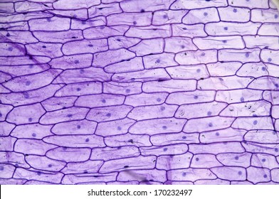
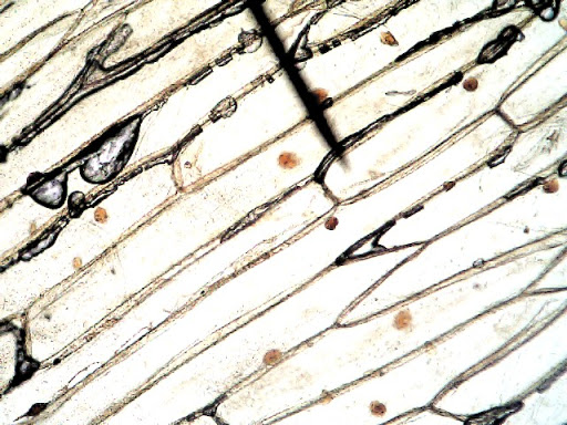
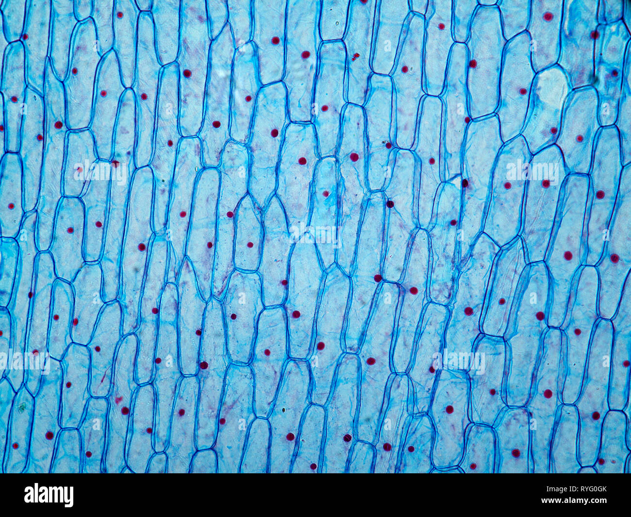
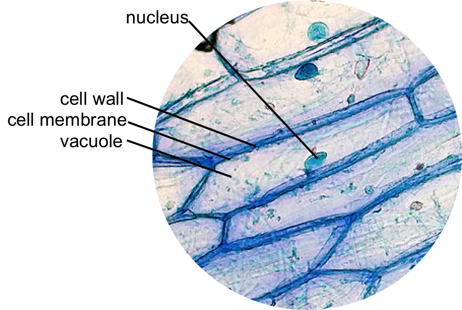
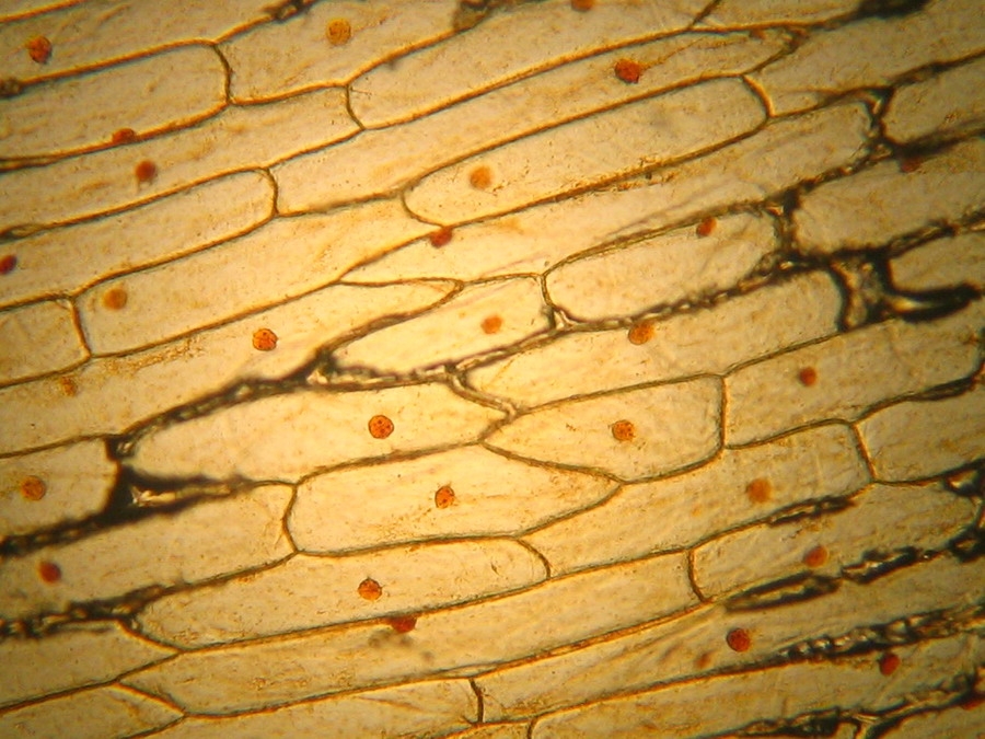
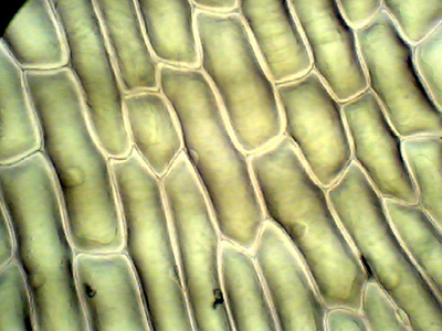
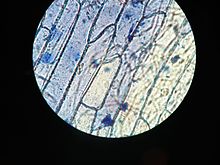
Onion Under Microscope. The cells look elongated similar in appearance color size and shape have thick cell walls and a nucleus that is large and circular in shape. There are different types of stains depending on what type of cell you are going to look at. You ll need to stain the onion cells before you observe them under the microscope. Take a piece from on of the sections and peel off a small thin piece of the onion epidermis or skin.
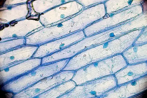 Onion Cells Under A Microscope Requirements Preparation Observation From microscopemaster.com
Root tip of onion and mitosis cell in the root tip for education. Onion cells under the microscope introduction the bulb of an onion is formed from modified leaves. While photosynthesis takes place in the leaves of an onion containing chloroplast the little glucose that is produced from this process is converted in to starch starch granules and stored in the bulb. Take a piece from on of the sections and peel off a small thin piece of the onion epidermis or skin. Although onions may not have as much starch as potato and other plants the stain iodine allows for the little starch molecules to be visible under the microscope. This is to hold the onion skin and to keep it from drying out.
You do not need to copy out this list of materials or the procedure below since it has already been done for you.
Onion cells under the microscope introduction the bulb of an onion is formed from modified leaves. A stereo microscope allows you to see the surface of specimens with a 3 dimensional view. Cut the onion into sections. The cells look elongated similar in appearance color size and shape have thick cell walls and a nucleus that is large and circular in shape. 1 compound light microscope. Although onions may not have as much starch as potato and other plants the stain iodine allows for the little starch molecules to be visible under the microscope.
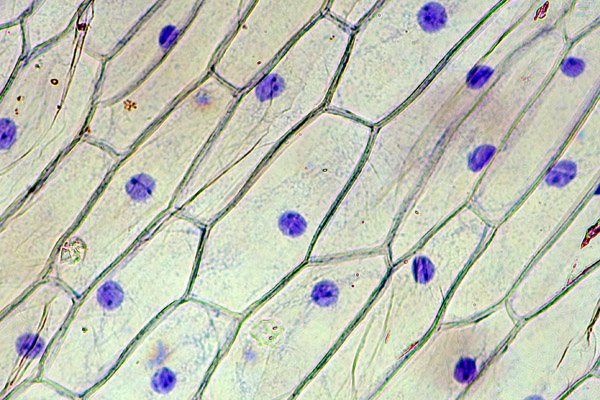 Source: microbehunter.com
You ll need to stain the onion cells before you observe them under the microscope. Although onions are plants students will not see any chloroplasts in their slides. Cut the onion into sections. 1 compound light microscope. The cells look elongated similar in appearance color size and shape have thick cell walls and a nucleus that is large and circular in shape.
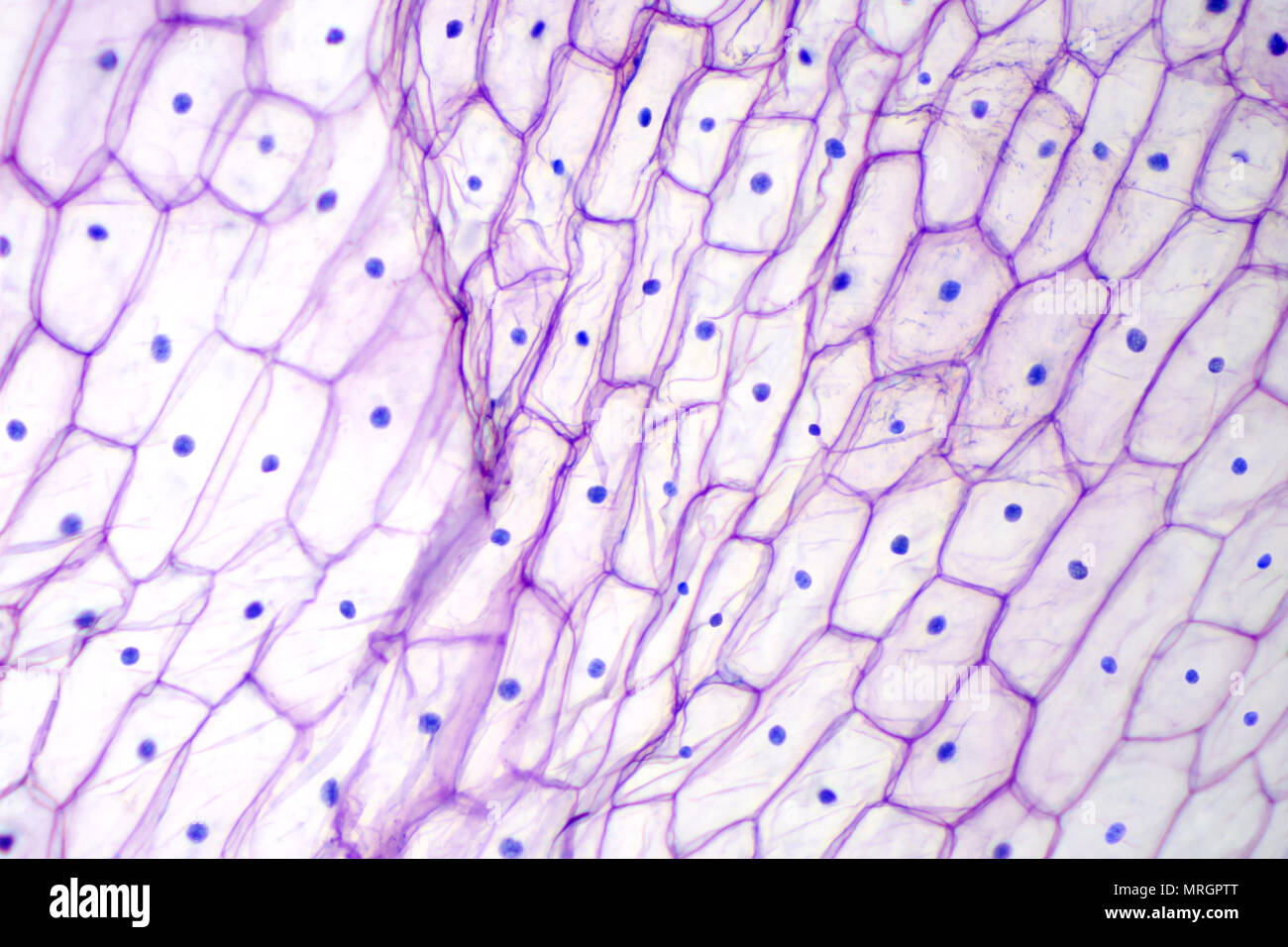 Source: alamy.com
1 slide 1 cover slip 1 pair of tweezers 1 eye dropper a few drops of water 1 paper towel 1 small piece of onion procedure. 1 compound light microscope. Onion cells under the microscope introduction the bulb of an onion is formed from modified leaves. You ll need to stain the onion cells before you observe them under the microscope. A stereo microscope allows you to see the surface of specimens with a 3 dimensional view.
 Source: shutterstock.com
You do not need to copy out this list of materials or the procedure below since it has already been done for you. The cells are easily visible under a microscope and the preparation of a thin section is straight forward. It provides some contrast for viewing under a microscope. Root tip of onion and mitosis cell in the root tip for education. A stereo microscope allows you to see the surface of specimens with a 3 dimensional view.
 Source: reddit.com
An onion is made of layers each separated by a thin skin or membrane. Iodine dark stain that colors starches in cells. Onion cells under microscope mitosis written by macpride friday july 17 2020 add comment edit. There are different types of stains depending on what type of cell you are going to look at. 1 slide 1 cover slip 1 pair of tweezers 1 eye dropper a few drops of water 1 paper towel 1 small piece of onion procedure.
 Source: m.youtube.com
1 slide 1 cover slip 1 pair of tweezers 1 eye dropper a few drops of water 1 paper towel 1 small piece of onion procedure. First place a small drop of water on a microscope slide. Under a stereo microscope you can see the metallic texture and colors of the mosquito s compound eyes. Root tip of onion and mitosis cell in the root tip of onion under. You ll need to stain the onion cells before you observe them under the microscope.
 Source: microscopemaster.com
Cells present in onion peel can be observed under microscope. Take a piece from on of the sections and peel off a small thin piece of the onion epidermis or skin. It provides some contrast for viewing under a microscope. First place a small drop of water on a microscope slide. 1 compound light microscope.
 Source: biology-pictures.blogspot.com
For this experiment outer most scale of the onion is removed and is cut into four equal halves. In contrast the light has to pass through the specimen to form the image under a compound microscope. Onion cells under microscope mitosis written by macpride friday july 17 2020 add comment edit. 1 slide 1 cover slip 1 pair of tweezers 1 eye dropper a few drops of water 1 paper towel 1 small piece of onion procedure. Take a piece from on of the sections and peel off a small thin piece of the onion epidermis or skin.
 Source: alamy.com
While photosynthesis takes place in the leaves of an onion containing chloroplast the little glucose that is produced from this process is converted in to starch starch granules and stored in the bulb. For this experiment outer most scale of the onion is removed and is cut into four equal halves. The cells are easily visible under a microscope and the preparation of a thin section is straight forward. Take a piece from on of the sections and peel off a small thin piece of the onion epidermis or skin. It is a monocot plant.
 Source: m.youtube.com
It is a monocot plant. Although onions may not have as much starch as potato and other plants the stain iodine allows for the little starch molecules to be visible under the microscope. Cut the onion into sections. Onion cells under the microscope introduction the bulb of an onion is formed from modified leaves. 1 slide 1 cover slip 1 pair of tweezers 1 eye dropper a few drops of water 1 paper towel 1 small piece of onion procedure.
 Source: saurabhg.com
While photosynthesis takes place in the leaves of an onion containing chloroplast the little glucose that is produced from this process is converted in to starch starch granules and stored in the bulb. 1 compound light microscope. An onion is made of layers each separated by a thin skin or membrane. Cells present in onion peel can be observed under microscope. A stereo microscope allows you to see the surface of specimens with a 3 dimensional view.
 Source: pinterest.com
Have one partner take a microscope from the back of the room. While photosynthesis takes place in the leaves of an onion containing chloroplast the little glucose that is produced from this process is converted in to starch starch granules and stored in the bulb. Tissue from an onion is a good first exercise in using the microscope and viewing plant cells. First place a small drop of water on a microscope slide. 1 slide 1 cover slip 1 pair of tweezers 1 eye dropper a few drops of water 1 paper towel 1 small piece of onion procedure.
 Source: microscopy-uk.org.uk
Iodine dark stain that colors starches in cells. Cells present in onion peel can be observed under microscope. It is a monocot plant. Cut the onion into sections. In contrast the light has to pass through the specimen to form the image under a compound microscope.
 Source: microscopemaster.com
It is a monocot plant. For this onion peels are first isolated. The cells look elongated similar in appearance color size and shape have thick cell walls and a nucleus that is large and circular in shape. Onion cells under the microscope introduction the bulb of an onion is formed from modified leaves. Cells present in onion peel can be observed under microscope.
 Source: en.wikibooks.org
Root tip of onion and mitosis cell in the root tip of onion under. An onion is made of layers each separated by a thin skin or membrane. Root tip of onion and mitosis cell in the root tip for education. First place a small drop of water on a microscope slide. Then with the help of a pairs of forcep the scale of onion is peeled out.
 Source: 123rf.com
It is a monocot plant. Iodine dark stain that colors starches in cells. While photosynthesis takes place in the leaves of an onion containing chloroplast the little glucose that is produced from this process is converted in to starch starch granules and stored in the bulb. Take a piece from on of the sections and peel off a small thin piece of the onion epidermis or skin. The main onion cell structures are quite easy to observe under medium magnification levels when using a light microscope.
If you find this site serviceableness, please support us by sharing this posts to your favorite social media accounts like Facebook, Instagram and so on or you can also bookmark this blog page with the title onion under microscope by using Ctrl + D for devices a laptop with a Windows operating system or Command + D for laptops with an Apple operating system. If you use a smartphone, you can also use the drawer menu of the browser you are using. Whether it’s a Windows, Mac, iOS or Android operating system, you will still be able to bookmark this website.


