Preparation of microscopic slide
Preparation Of Microscopic Slide. This is a collection of aerobiology and mold testing lab standard operating procedures sop s and slide preparation recipes useful for identification of mold pollen animal dander skin cells mite fecals mouse dander and other airborne allergens and bioaerosols. How to prepare microscope slides for lab analysis. Dry mounts wet mount squash staining. Clipping is a handy way to collect important slides you want to.
 Slide Preparation L O To Be Able To Prepare A Microscope Slide Ppt Video Online Download From slideplayer.com
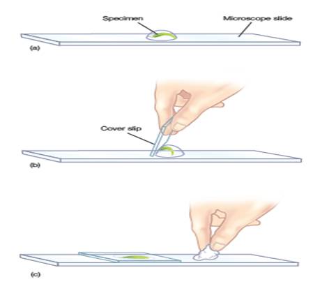
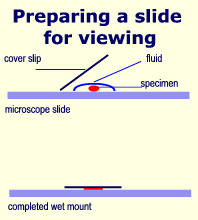
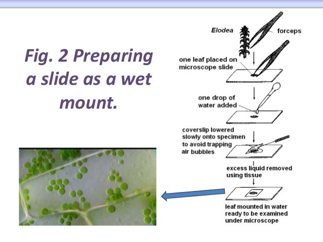
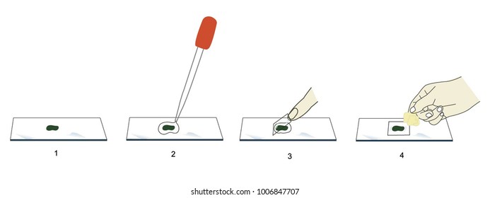
Dry mounts wet mount squash staining. Standard techniques such as wet mounting dry mounting and smearing are used under certain circumstances to observe specimens under a microscope. A small square of clear glass or plastic a coverslip is placed on top of the liquid to minimize evaporation and protect the microscope lens from exposure to the sample. You just clipped your first slide. How to prepare microscope slides for lab analysis. Cover it with a coverslip with the help of needle.
To prepare a wet mount using a flat slide or a depression slide.
Preparation of specimen for microscopic examination 1. Dry the slide using a clean cloth. The main methods of placing samples onto microscope slides are wet mount dry mount smear squash and staining. Place a drop of fluid in the middle of the slide e g water glycerin immersion oil or a liquid sample. The preparation of a microscopic slide may take hours and for those who have no experience the slide may not be well prepared for viewing which is time wasted. Simply position a thinly sliced section on the center of the slide and place a cover slip over the sample.
 Source: thoughtco.com
Focus the slide under lower of microscope and then change to high power if needed precautions. Simply position a thinly sliced section on the center of the slide and place a cover slip over the sample. If you find that your microscope slide has any contamination on it including your own fingerprints give it a quick wash with liquid soap and water. Clipping is a handy way to collect important slides you want to. The main methods of placing samples onto microscope slides are wet mount dry mount smear squash and staining.
 Source: academygenbioii.pbworks.com
The main methods of placing samples onto microscope slides are wet mount dry mount smear squash and staining. This is a collection of aerobiology and mold testing lab standard operating procedures sop s and slide preparation recipes useful for identification of mold pollen animal dander skin cells mite fecals mouse dander and other airborne allergens and bioaerosols. Observe it under a compound microscope after staining and mounting. Clipping is a handy way to collect important slides you want to. The preparation of a microscopic slide may take hours and for those who have no experience the slide may not be well prepared for viewing which is time wasted.
 Source: phywe.com
The dry mount is the most basic technique. Preparation of specimen for microscopic examination deep patel. The main methods of placing samples onto microscope slides are wet mount dry mount smear squash and staining. Observe it under a compound microscope after staining and mounting. The dry mount is the most basic technique.
 Source: irevise.com
If you find that your microscope slide has any contamination on it including your own fingerprints give it a quick wash with liquid soap and water. Do not use tissues or paper towels as these can leave lint behind. The dry mount is the most basic technique. Observe it under a compound microscope after staining and mounting. The main methods of placing samples onto microscope slides are wet mount dry mount smear squash and staining.
 Source: fishdoc.co.uk
Microscope slide preparation is the process of readying a slide for observation under an optical microscope. The preparation of a microscopic slide may take hours and for those who have no experience the slide may not be well prepared for viewing which is time wasted. Put the thinnest section in the centre of the slide. Dry mounts wet mount squash staining. A small square of clear glass or plastic a coverslip is placed on top of the liquid to minimize evaporation and protect the microscope lens from exposure to the sample.
 Source: youtube.com
How to prepare microscope slides for lab analysis. Cover it with a coverslip with the help of needle. Aerobiolgy forensic microscopy lab procedures. A small square of clear glass or plastic a coverslip is placed on top of the liquid to minimize evaporation and protect the microscope lens from exposure to the sample. You just clipped your first slide.
 Source: en.wikibooks.org
This is a collection of aerobiology and mold testing lab standard operating procedures sop s and slide preparation recipes useful for identification of mold pollen animal dander skin cells mite fecals mouse dander and other airborne allergens and bioaerosols. Preparation of specimen for microscopic examination 1. This is a collection of aerobiology and mold testing lab standard operating procedures sop s and slide preparation recipes useful for identification of mold pollen animal dander skin cells mite fecals mouse dander and other airborne allergens and bioaerosols. Standard techniques such as wet mounting dry mounting and smearing are used under certain circumstances to observe specimens under a microscope. Place a drop of fluid in the middle of the slide e g water glycerin immersion oil or a liquid sample.
 Source: microscopegenius.com
Observe it under a compound microscope after staining and mounting. Dry mounts wet mount squash staining. If you find that your microscope slide has any contamination on it including your own fingerprints give it a quick wash with liquid soap and water. Put the thinnest section in the centre of the slide. Preparation of specimen for microscopic examination 1.
 Source: microscope-microscope.org
Simply position a thinly sliced section on the center of the slide and place a cover slip over the sample. This is a collection of aerobiology and mold testing lab standard operating procedures sop s and slide preparation recipes useful for identification of mold pollen animal dander skin cells mite fecals mouse dander and other airborne allergens and bioaerosols. The main methods of placing samples onto microscope slides are wet mount dry mount smear squash and staining. Tech ii iv semester materials science metallurgy department of mechanical engineering. To prepare a wet mount using a flat slide or a depression slide.
 Source: researchgate.net
Study under the microscope. Do not use tissues or paper towels as these can leave lint behind. Place a drop of fluid in the middle of the slide e g water glycerin immersion oil or a liquid sample. Put the thinnest section in the centre of the slide. Focus the slide under lower of microscope and then change to high power if needed precautions.
 Source: slideshare.net
You just clipped your first slide. A small square of clear glass or plastic a coverslip is placed on top of the liquid to minimize evaporation and protect the microscope lens from exposure to the sample. To prepare a wet mount using a flat slide or a depression slide. Tech ii iv semester materials science metallurgy department of mechanical engineering. Observe it under a compound microscope after staining and mounting.
 Source: wikihow.com
Tech ii iv semester materials science metallurgy department of mechanical engineering. A small square of clear glass or plastic a coverslip is placed on top of the liquid to minimize evaporation and protect the microscope lens from exposure to the sample. Moreover the prepared microscope slide set is composed of a good number of preparations given that it contains 50 to 100 slides. Simply position a thinly sliced section on the center of the slide and place a cover slip over the sample. You just clipped your first slide.
 Source: slideplayer.com
Moreover the prepared microscope slide set is composed of a good number of preparations given that it contains 50 to 100 slides. Dry mounts wet mount squash staining. You just clipped your first slide. Do not use tissues or paper towels as these can leave lint behind. How to prepare microscope slides for lab analysis.
 Source: shutterstock.com
Dry mounts wet mount squash staining. Standard techniques such as wet mounting dry mounting and smearing are used under certain circumstances to observe specimens under a microscope. Preparation of specimen for microscopic examination deep patel. The main methods of placing samples onto microscope slides are wet mount dry mount smear squash and staining. The dry mount is the most basic technique.
 Source: microscopy-uk.org.uk
Do not use tissues or paper towels as these can leave lint behind. A small square of clear glass or plastic a coverslip is placed on top of the liquid to minimize evaporation and protect the microscope lens from exposure to the sample. Clipping is a handy way to collect important slides you want to. Moreover the prepared microscope slide set is composed of a good number of preparations given that it contains 50 to 100 slides. If you find that your microscope slide has any contamination on it including your own fingerprints give it a quick wash with liquid soap and water.
If you find this site convienient, please support us by sharing this posts to your preference social media accounts like Facebook, Instagram and so on or you can also bookmark this blog page with the title preparation of microscopic slide by using Ctrl + D for devices a laptop with a Windows operating system or Command + D for laptops with an Apple operating system. If you use a smartphone, you can also use the drawer menu of the browser you are using. Whether it’s a Windows, Mac, iOS or Android operating system, you will still be able to bookmark this website.

