Sheep brain dissection lab
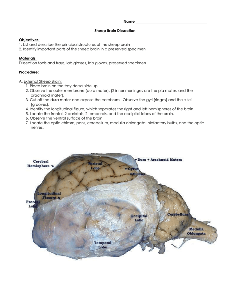
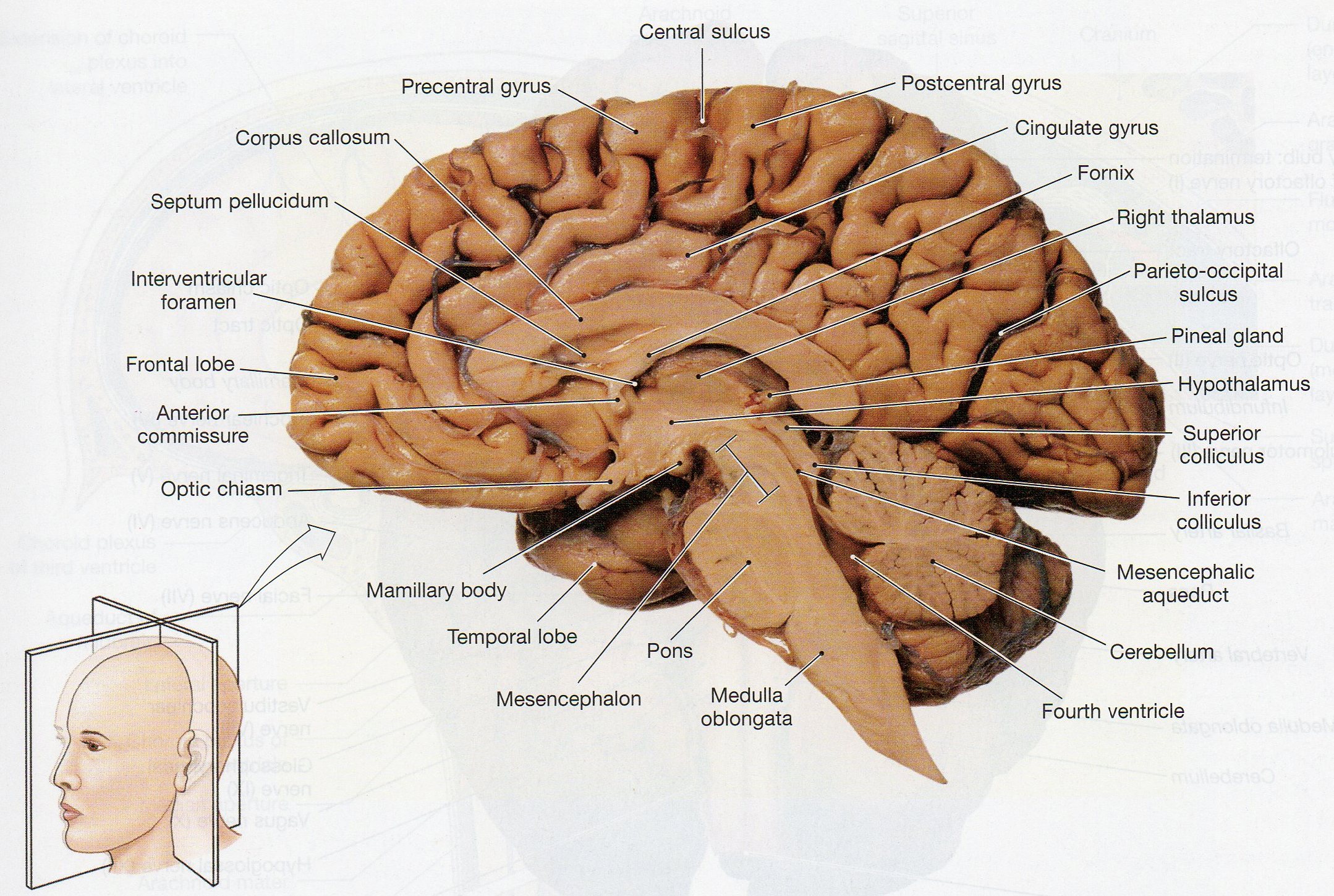
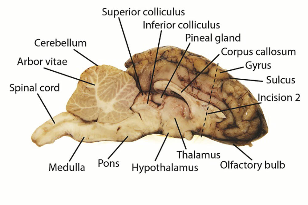
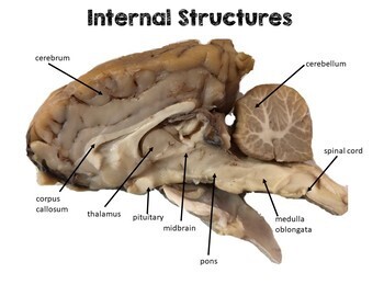
Sheep Brain Dissection Lab. Refer to images descriptions and functions of parts of the brain as you proceed through this lab. During this lab i cut open a sheep brain and labeled all the divided structures that allow a sheep to survive and sustain life when everything worked as one. 11 the image below shows a cleanly separated brain with the major internal structures visible and labeled. Dissection tools and trays lab glasses lab gloves preserved specimen.
 Sheep Brain Dissection From studylib.net
Examine the ventral surface of the sheep brain. Muscles other nerves and fatty. With each type of brain there will be differences in size structure and brain regions but the general structures and their area of location are relatively the same or very similar. A pair of olfactory bulbs may be seen one under each lobe of the frontal cortex. By dissecting and labeling the sheep brain and cow. The sheep brain is quite similar to the human brain except for proportion.
1 sheep brain and cow eye dissection lab report ivy tech anatomy and physiology 101 2 27 2020 abstract the purpose of the sheep brain and cow eye dissection is to familiarize locating and identify the regions and structures in the brain and eye.
Dissection tools and trays lab glasses lab gloves preserved specimen. Sheep brain dissection lab guide sheep brain labeling cranial nerves coloring gallery of dissection images. External anatomy of sheep brain. The sheep brain and cow eye were used because their functions are similar of a human brain and eye. Several important parts of the visual system are visible in the ventral view of the brain. The sheep brain is quite similar to the human brain except for proportion.
 Source: chegg.com
1 sheep brain and cow eye dissection lab report ivy tech anatomy and physiology 101 2 27 2020 abstract the purpose of the sheep brain and cow eye dissection is to familiarize locating and identify the regions and structures in the brain and eye. You ll need a preserved sheep brain for the dissection. Below is a brief survey of the internal and external anatomy of the sheep brain. Dissection tools and trays lab glasses lab gloves preserved specimen. As students dissect a sheep brain they ll even gain a better understanding of.
 Source: pinterest.com
The sheep brain and cow eye were used because their functions are similar of a human brain and eye. Several important parts of the visual system are visible in the ventral view of the brain. 1 sheep brain and cow eye dissection lab report ivy tech anatomy and physiology 101 2 27 2020 abstract the purpose of the sheep brain and cow eye dissection is to familiarize locating and identify the regions and structures in the brain and eye. The sheep brain is quite similar to the human brain except for proportion. The sheep brain is quite similar to the human brain except for proportion.
Source:
During this lab i cut open a sheep brain and labeled all the divided structures that allow a sheep to survive and sustain life when everything worked as one. The sheep brain and cow eye were used because their functions are similar of a human brain and eye. Closely examine this sheep organ to learn about structures of the brain such as the cerebellum cranial nerve and so much more. 11 the image below shows a cleanly separated brain with the major internal structures visible and labeled. A pair of olfactory bulbs may be seen one under each lobe of the frontal cortex.
 Source: courses.lumenlearning.com
With each type of brain there will be differences in size structure and brain regions but the general structures and their area of location are relatively the same or very similar. Dissection tools and tray lab gloves preserved sheep. Refer to images descriptions and functions of parts of the brain as you proceed through this lab. With each type of brain there will be differences in size structure and brain regions but the general structures and their area of location are relatively the same or very similar. Below is a brief survey of the internal and external anatomy of the sheep brain.
 Source: learning-center.homesciencetools.com
Sheep brain dissection lab guide sheep brain labeling cranial nerves coloring gallery of dissection images. Examine the ventral surface of the sheep brain. The sheep brain is quite similar to the human brain except for proportion. The sheep brain is quite similar to the human brain except for proportion. The sheep has a smaller cerebrum.
 Source: youtube.com
Sheep brain dissection 1 before starting this lab open the brain parts and functions document. Sheep brain dissection lab guide sheep brain labeling cranial nerves coloring gallery of dissection images. Also the sheep brain is oriented anterior to posterior more horizontally while the human brain is oriented superior to interior more vertically materials. With each type of brain there will be differences in size structure and brain regions but the general structures and their area of location are relatively the same or very similar. The sheep brain and cow eye were used because their functions are similar of a human brain and eye.
 Source: www2.palomar.edu
The sheep brain is quite similar to the human brain except for proportion. Also the sheep brain is oriented anterior to posterior whereas the human brain is superior to inferior. By dissecting and labeling the sheep brain and cow. Closely examine this sheep organ to learn about structures of the brain such as the cerebellum cranial nerve and so much more. Refer to images descriptions and functions of parts of the brain as you proceed through this lab.
 Source: studylib.net
A pair of olfactory bulbs may be seen one under each lobe of the frontal cortex. Also the sheep brain is oriented anterior to posterior more horizontally while the human brain is oriented superior to interior more vertically materials. Sheep brain dissection 1 before starting this lab open the brain parts and functions document. The sheep brain and cow eye were used because their functions are similar of a human brain and eye. During this lab i cut open a sheep brain and labeled all the divided structures that allow a sheep to survive and sustain life when everything worked as one.
 Source: biology4friends.org
You ll need a preserved sheep brain for the dissection. You ll need a preserved sheep brain for the dissection. Refer to images descriptions and functions of parts of the brain as you proceed through this lab. The next several steps will view this surface of the brain. As students dissect a sheep brain they ll even gain a better understanding of.
 Source: starpathdesign.com
Several important parts of the visual system are visible in the ventral view of the brain. A preserved sheep brain specimen a photographic sheep brain dissection guide a 22 scalpel a magnifying glass and a dissecting tray. Use this for a high school lab or just look at the labeled images to get an idea of what the brain looks like. The sheep brain is quite similar to the human brain except for proportion. Carolina s perfect solution sheep brain dissection introduces students to the anatomy of a mammalian brain both external and internal and encourages students to construct an explanation of the central nervous system.
 Source: learning-center.homesciencetools.com
A pair of olfactory bulbs may be seen one under each lobe of the frontal cortex. Finally a section of the brain is cut to examine the difference between white matter and gray matter. Several important parts of the visual system are visible in the ventral view of the brain. The sheep has a smaller cerebrum. Carolina s perfect solution sheep brain dissection introduces students to the anatomy of a mammalian brain both external and internal and encourages students to construct an explanation of the central nervous system.
 Source: carolina.com
Sheep brain dissection lab guide sheep brain labeling cranial nerves coloring gallery of dissection images. Sheep brain dissection guide 3. The next several steps will view this surface of the brain. Also the sheep brain is oriented anterior to posterior more horizontally while the human brain is oriented superior to interior more vertically materials. A preserved sheep brain specimen a photographic sheep brain dissection guide a 22 scalpel a magnifying glass and a dissecting tray.
 Source: jb004.k12.sd.us
11 the image below shows a cleanly separated brain with the major internal structures visible and labeled. Dissection tools and tray lab gloves preserved sheep. A pair of olfactory bulbs may be seen one under each lobe of the frontal cortex. Refer to images descriptions and functions of parts of the brain as you proceed through this lab. Carolina s perfect solution sheep brain dissection introduces students to the anatomy of a mammalian brain both external and internal and encourages students to construct an explanation of the central nervous system.
 Source: biologycorner.com
The sheep brain is quite similar to the human brain except for proportion. Below is a brief survey of the internal and external anatomy of the sheep brain. Several important parts of the visual system are visible in the ventral view of the brain. Sheep brains although much smaller than human brains have similar features and can be a valuable addition to anatomy studies. Dissection tools and trays lab glasses lab gloves preserved specimen.
 Source: teacherspayteachers.com
11 the image below shows a cleanly separated brain with the major internal structures visible and labeled. The sheep has a smaller cerebrum. The sheep brain is quite similar to the human brain except for proportion. External anatomy of sheep brain. With each type of brain there will be differences in size structure and brain regions but the general structures and their area of location are relatively the same or very similar.
If you find this site beneficial, please support us by sharing this posts to your favorite social media accounts like Facebook, Instagram and so on or you can also bookmark this blog page with the title sheep brain dissection lab by using Ctrl + D for devices a laptop with a Windows operating system or Command + D for laptops with an Apple operating system. If you use a smartphone, you can also use the drawer menu of the browser you are using. Whether it’s a Windows, Mac, iOS or Android operating system, you will still be able to bookmark this website.


