Sheep brain dissection pictures
Sheep Brain Dissection Pictures. The sheep brain is exposed and each of the structures are labeled and described in a sequential manner in the same way that a real dissection would occur. You ll need a preserved sheep brain for the dissection. Learn sheep brain pictures with free interactive flashcards. Muscles other nerves and fatty.
 Dr Parker S A P I Sheep Brain Dissection Youtube From youtube.com
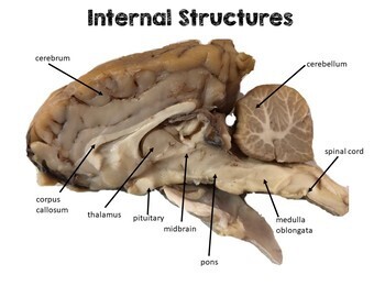
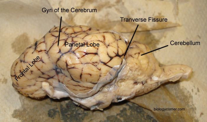
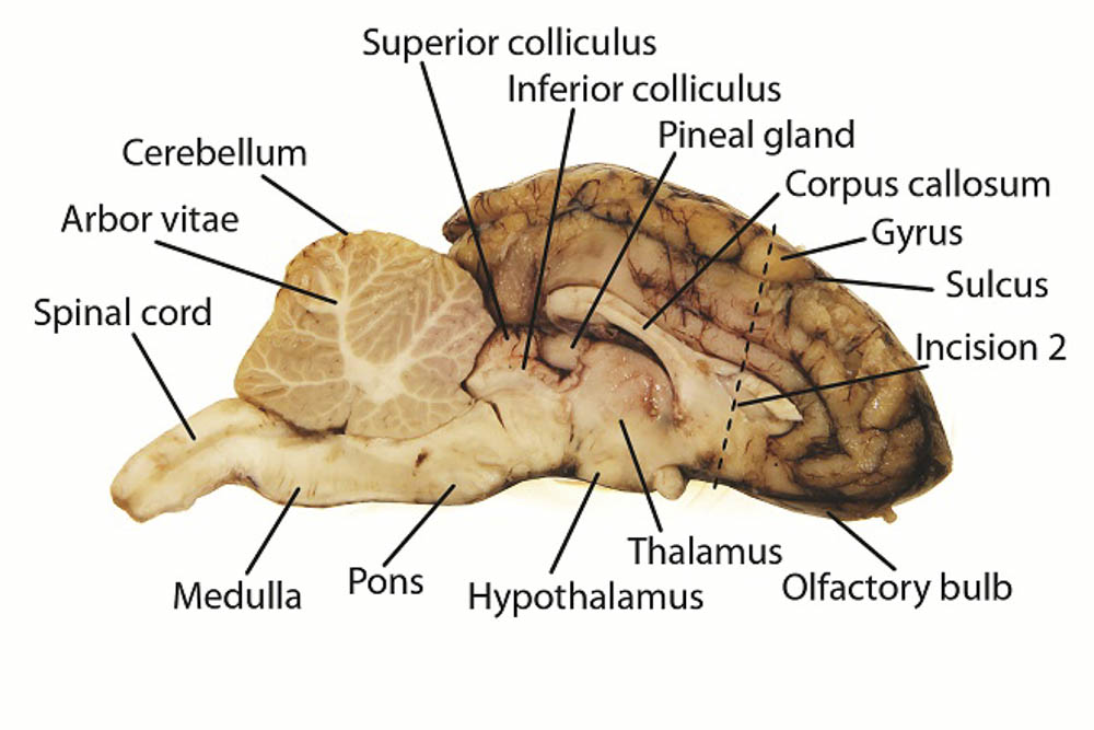
See for yourself what the cerebrum cerebellum spinal cord gray and white matter and other parts of the brain look like. Set the brain down so the flatter side with the white spinal cord at one end rests on the dissection pan. Use this for a high school lab or just look at the labeled images to get an idea of what the brain looks like. Images of parts of a sheep brain. Learn sheep brain pictures with free interactive flashcards. This shows the ventral side of the brain with the infundibulum optic chiasma pons and medulla oblongata visible.
Obtain a sheep brain dissection styrofoam tray gloves and surgical tools take pictures before and after each step of removal cutting or labeling.
A pair of olfactory bulbs may be seen one under each lobe of the frontal cortex. Follow the list of structures the professor outlines and identify by labeling 80 of them on the. The next several steps will view this surface of the brain. Muscles other nerves and fatty. The sheep brain is exposed and each of the structures are labeled and described in a sequential manner in the same way that a real dissection would occur. This shows the ventral side of the brain with the infundibulum optic chiasma pons and medulla oblongata visible.
 Source: teacherspayteachers.com
Gallery of dissection images. Choose from 500 different sets of sheep brain pictures flashcards on quizlet. Muscles other nerves and fatty. Students dissected the brain. Recorded at glen oaks community college centreville michigan by dr ren allen hartung.
 Source: biologycorner.com
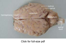
External anatomy of sheep brain. Learn sheep brain pictures with free interactive flashcards. Powered by create your own unique website with customizable templates. Recorded at glen oaks community college centreville michigan by dr ren allen hartung. The sheep brain is enclosed in a tough outer covering called the dura mater.
 Source: learning-center.homesciencetools.com
Students dissected the brain. Sheep brain dissection guide with pictures december 12 2016 sheep brains although much smaller than human brains have similar features and can be a valuable addition to anatomy studies. This is the url to a sheep dissection lab created by five students inthe bi college haverford and bryn mawr community. It is a fantastic siteto explore the brain. The next several steps will view this surface of the brain.
 Source: biology4friends.org
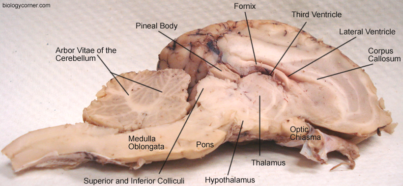
Cut the brain along midsagittal line and through the longitudinal fissure. Follow the list of structures the professor outlines and identify by labeling 80 of them on the. The sheep brain is enclosed in a tough outer covering called the dura mater. Powered by create your own unique website with customizable templates. This is the url to a sheep dissection lab created by five students inthe bi college haverford and bryn mawr community.
 Source: courses.lumenlearning.com
Set the brain down so the flatter side with the white spinal cord at one end rests on the dissection pan. The next several steps will view this surface of the brain. Jessica kuhn jessica magid karen revere elizabeth caris and gray vargasthrough science education at bryn mawr college. Learn sheep brain pictures with free interactive flashcards. The sheep brain is exposed and each of the structures are labeled and described in a sequential manner in the same way that a real dissection would occur.
 Source: pinterest.com
Use this for a high school lab or just look at the labeled images to get an idea of what the brain looks like. Choose from 500 different sets of sheep brain pictures flashcards on quizlet. You ll need a preserved sheep brain for the dissection. This is the url to a sheep dissection lab created by five students inthe bi college haverford and bryn mawr community. Use this for a high school lab or just look at the labeled images to get an idea of what the brain looks like.
 Source: m.youtube.com
Follow the list of structures the professor outlines and identify by labeling 80 of them on the. Choose from 500 different sets of sheep brain pictures flashcards on quizlet. Images of parts of a sheep brain. See for yourself what the cerebrum cerebellum spinal cord gray and white matter and other parts of the brain look like. Gallery of dissection images.
 Source: ihsvoice.com
Use this for a high school lab or just look at the labeled images to get an idea of what the brain looks like. The sheep brain is exposed and each of the structures are labeled and described in a sequential manner in the same way that a real dissection would occur. Obtain a sheep brain dissection styrofoam tray gloves and surgical tools take pictures before and after each step of removal cutting or labeling. Gallery of dissection images. Examine the ventral surface of the sheep brain.
 Source: m.carolina.com
Sheep brain dissection guide with pictures december 12 2016 sheep brains although much smaller than human brains have similar features and can be a valuable addition to anatomy studies. Powered by create your own unique website with customizable templates. The sheep brain is enclosed in a tough outer covering called the dura mater. It is a fantastic siteto explore the brain. See for yourself what the cerebrum cerebellum spinal cord gray and white matter and other parts of the brain look like.
 Source: youtube.com
The sheep brain is exposed and each of the structures are labeled and described in a sequential manner in the same way that a real dissection would occur. Several important parts of the visual system are visible in the ventral view of the brain. Powered by create your own unique website with customizable templates. Set the brain down so the flatter side with the white spinal cord at one end rests on the dissection pan. See for yourself what the cerebrum cerebellum spinal cord gray and white matter and other parts of the brain look like.
 Source: pinterest.ca
Cut the brain along midsagittal line and through the longitudinal fissure. You ll need a preserved sheep brain for the dissection. Students dissected the brain. Use this for a high school lab or just look at the labeled images to get an idea of what the brain looks like. The sheep brain is enclosed in a tough outer covering called the dura mater.
 Source: brainu.org
The sheep brain is enclosed in a tough outer covering called the dura mater. Set the brain down so the flatter side with the white spinal cord at one end rests on the dissection pan. Examine the ventral surface of the sheep brain. Obtain a sheep brain dissection styrofoam tray gloves and surgical tools take pictures before and after each step of removal cutting or labeling. Recorded at glen oaks community college centreville michigan by dr ren allen hartung.
 Source: jb004.k12.sd.us
Follow the list of structures the professor outlines and identify by labeling 80 of them on the. This shows the ventral side of the brain with the infundibulum optic chiasma pons and medulla oblongata visible. This is the url to a sheep dissection lab created by five students inthe bi college haverford and bryn mawr community. Sheep brain dissection guide with pictures december 12 2016 sheep brains although much smaller than human brains have similar features and can be a valuable addition to anatomy studies. The next several steps will view this surface of the brain.
 Source: biologycorner.com
Recorded at glen oaks community college centreville michigan by dr ren allen hartung. Follow the list of structures the professor outlines and identify by labeling 80 of them on the. Powered by create your own unique website with customizable templates. Set the brain down so the flatter side with the white spinal cord at one end rests on the dissection pan. External anatomy of sheep brain.
 Source: learning-center.homesciencetools.com
This shows the ventral side of the brain with the infundibulum optic chiasma pons and medulla oblongata visible. Cut the brain along midsagittal line and through the longitudinal fissure. Use this for a high school lab or just look at the labeled images to get an idea of what the brain looks like. Obtain a sheep brain dissection styrofoam tray gloves and surgical tools take pictures before and after each step of removal cutting or labeling. Recorded at glen oaks community college centreville michigan by dr ren allen hartung.
If you find this site adventageous, please support us by sharing this posts to your favorite social media accounts like Facebook, Instagram and so on or you can also bookmark this blog page with the title sheep brain dissection pictures by using Ctrl + D for devices a laptop with a Windows operating system or Command + D for laptops with an Apple operating system. If you use a smartphone, you can also use the drawer menu of the browser you are using. Whether it’s a Windows, Mac, iOS or Android operating system, you will still be able to bookmark this website.


