The choroid of the eye
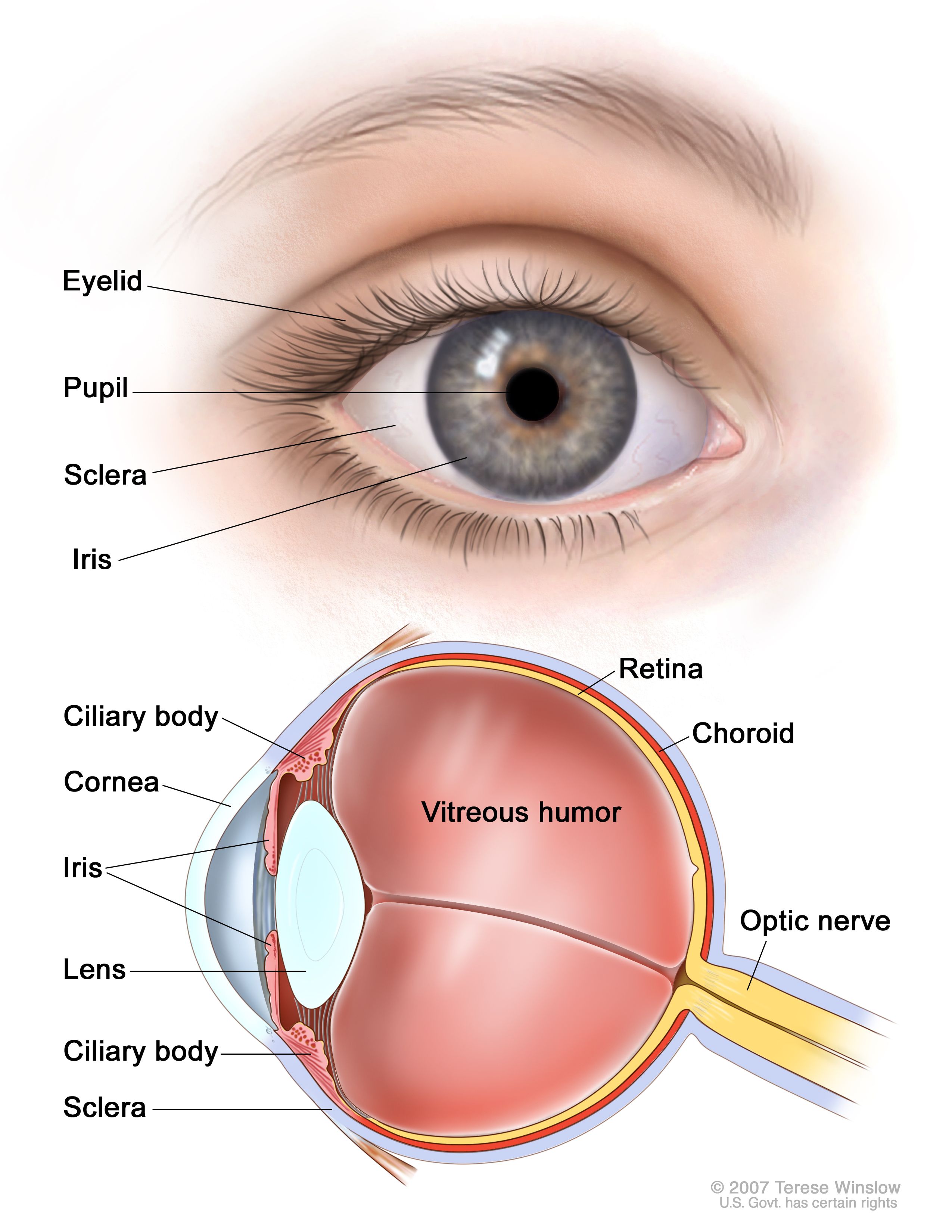
The Choroid Of The Eye. The choroid is the middle layer of the eye that contains blood vessels and connective tissue between the sclera the white of the eye and the retina at the back of the eye. The liver is the most common site for metastasis. Choroid is part of the uvea and supplies nutrients to the inner parts of the eye. Development of the lens v.
 Choroidal Neovascularization Wikipedia From en.wikipedia.org
Its function is to provide nourishment to the outer layers of the retina through blood vessels. The part of your eye between the sclera and the retina. Choroid is part of the uvea and supplies nutrients to the inner parts of the eye. By victoria ort ph d and david howard m d. Within this section of the eye there are four different layers. Development of the eye.
By victoria ort ph d and david howard m d.
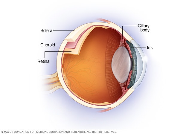
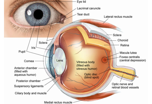
The human choroid is thickest at the far extreme rear of the eye while in the outlying areas it narrows to 0 1 mm. The choroid also known as the choroidea or choroid coat is the vascular layer of the eye containing connective tissues and lying between the retina and the sclera. The choroid provides oxygen and nourishment to the outer layers of the retina. The liver is the most common site for metastasis. The choroid is part of the uvea and it contains blood vessels and connective tissue. Development of the choroid sclera and cornea vi.
 Source: areaoftalmologica.com
Development of the iris and ciliary body vii. The choroid also known as the choroidea or choroid coat is the vascular layer of the eye containing connective tissues and lying between the retina and the sclera. It is delineated from the anterior part of the uvea called the ciliary body at the ora serrata 1. Eyelid and conjunctiva ix. Within this section of the eye there are four different layers.
 Source: cancer.gov
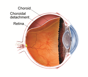
Choroid is part of the uvea and supplies nutrients to the inner parts of the eye. 1 it contains the retinal pigmented epithelial cells and provides oxygen and nourishment to the outer retina. The choroid also known as the choroid coat or choroidea is located between the retina and sclera. It joins the ciliary body anteriorly and lies between the retina and sclera posteriorly. Over time many choroidal melanomas enlarge and cause the retina to detach.
 Source: en.wikipedia.org
The choroid also known as the choroidea or choroid coat is the vascular layer of the eye containing connective tissues and lying between the retina and the sclera. This can lead to vision loss. Development of the retina iv. Development of the lens v. The choroid is the vascular layer of the eye that lies between the retina and the sclera.
 Source: mayoclinic.org
The function of the choroid is to provide oxygen and nutrients to the outer layers of the retina 2. The choroid is the vascular layer of the eye that lies between the retina and the sclera. The choroid also known as the choroid coat or choroidea is located between the retina and sclera. Its main purpose is to send oxygen and other nutrients to the retina. This can lead to vision loss.
 Source: pceyeglasses.com
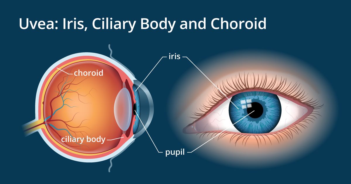
Over time many choroidal melanomas enlarge and cause the retina to detach. The vascular major blood vessel central layer of the eye lying between the retina and sclera. Along with the ciliary body and iris the choroid forms the uveal tract. Its function is to provide nourishment to the outer layers of the retina through blood vessels. My dashboard my education find an ophthalmologist.
 Source: allaboutvision.com
Choroid is part of the uvea and supplies nutrients to the inner parts of the eye. The vascular major blood vessel central layer of the eye lying between the retina and sclera. The choroid is part of the uvea and it contains blood vessels and connective tissue. The choroid is rich in blood vessels and supplies nutrients to the retina. The choroid is the middle layer of the eye that contains blood vessels and connective tissue between the sclera the white of the eye and the retina at the back of the eye.
 Source: researchgate.net
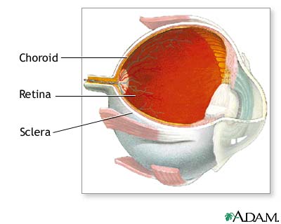
The choroid provides oxygen and nourishment to the outer layers of the retina. My dashboard my education find an ophthalmologist. The liver is the most common site for metastasis. The choroid is the vascular layer of the eye that lies between the retina and the sclera. The choroid is a thin variably pigmented vascular tissue forming the posterior uvea.
 Source: fightingblindness.ie
Development of the retina iv. The liver is the most common site for metastasis. Development of the lens v. The choroid is a thin variably pigmented vascular tissue forming the posterior uvea. Development of the iris and ciliary body vii.
 Source: medlineplus.gov
The choroid is the vascular layer of the eye that lies between the retina and the sclera. Development of the lens v. The choroid is a thin variably pigmented vascular tissue forming the posterior uvea. Inflammation of the choroid is called choroiditis. Development of the choroid sclera and cornea vi.
 Source: associatedretinaconsultants.com
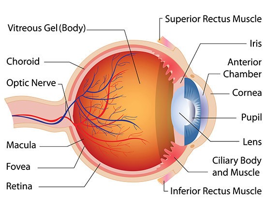
Development of the lens v. Development of the lens v. The choroid also known as the choroid coat or choroidea is located between the retina and sclera. Within this section of the eye there are four different layers. The choroid is extremely vascular with its capillaries arranged in a single layer on the inner surface to nourish the outer retinal layers figure 11 14.
 Source: asrs.org
The choroid is the vascular layer of the eye that lies between the retina and the sclera. It joins the ciliary body anteriorly and lies between the retina and sclera posteriorly. Development of the lens v. The choroid also known as the choroidea or choroid coat is the vascular layer of the eye containing connective tissues and lying between the retina and the sclera. Technically the choroid is the vascular part of the human eye that includes the connective tissue.
 Source: provisu.ch
The liver is the most common site for metastasis. In species with limited retinal vasculature e g horse rabbit guinea pig the retina depends to a large extent on the choroidal blood supply. Its function is to provide nourishment to the outer layers of the retina through blood vessels. The liver is the most common site for metastasis. The vascular major blood vessel central layer of the eye lying between the retina and sclera.
 Source: pinterest.com
The choroid is a thin variably pigmented vascular tissue forming the posterior uvea. The choroid also known as the choroid coat or choroidea is located between the retina and sclera. The human choroid is thickest at the far extreme rear of the eye while in the outlying areas it narrows to 0 1 mm. Over time many choroidal melanomas enlarge and cause the retina to detach. The part of your eye between the sclera and the retina.
Source: aao.org
It joins the ciliary body anteriorly and lies between the retina and sclera posteriorly. The choroid is the middle layer of the eye that contains blood vessels and connective tissue between the sclera the white of the eye and the retina at the back of the eye. The human choroid is thickest at the far extreme rear of the eye while in the outlying areas it narrows to 0 1 mm. Technically the choroid is the vascular part of the human eye that includes the connective tissue. Within this section of the eye there are four different layers.
 Source: en.wikipedia.org
The choroid is part of the uvea and it contains blood vessels and connective tissue. Development of the optic cup and lens vesicle iii. The choroid is part of the uvea and it contains blood vessels and connective tissue. Choroid is part of the uvea and supplies nutrients to the inner parts of the eye. Development of the retina iv.
If you find this site value, please support us by sharing this posts to your favorite social media accounts like Facebook, Instagram and so on or you can also save this blog page with the title the choroid of the eye by using Ctrl + D for devices a laptop with a Windows operating system or Command + D for laptops with an Apple operating system. If you use a smartphone, you can also use the drawer menu of the browser you are using. Whether it’s a Windows, Mac, iOS or Android operating system, you will still be able to bookmark this website.

