Where is the choroid located in the eye
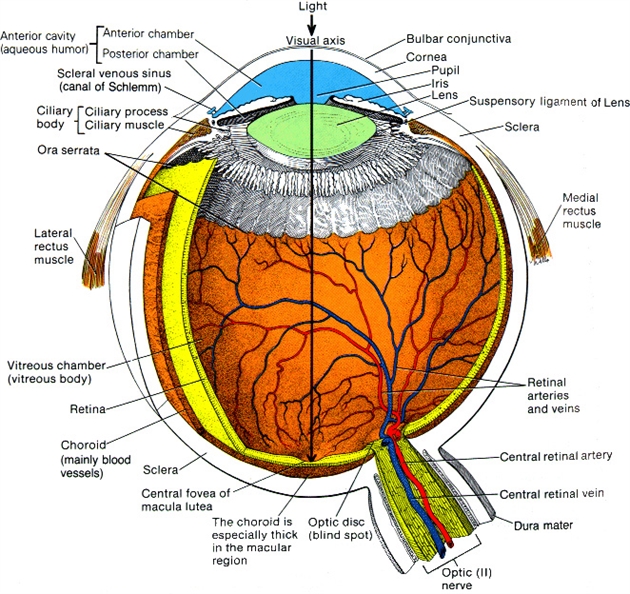
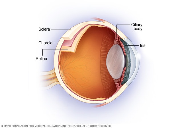
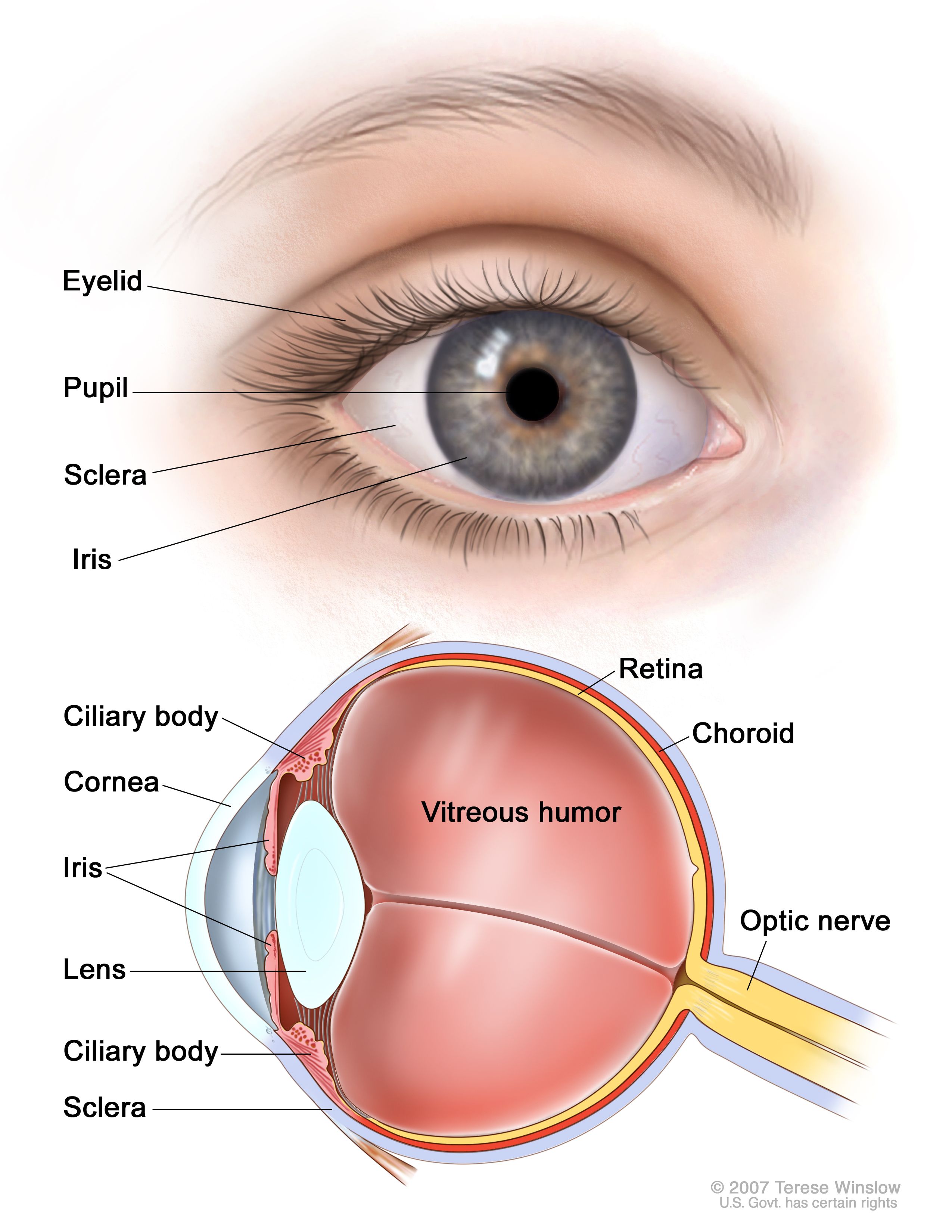
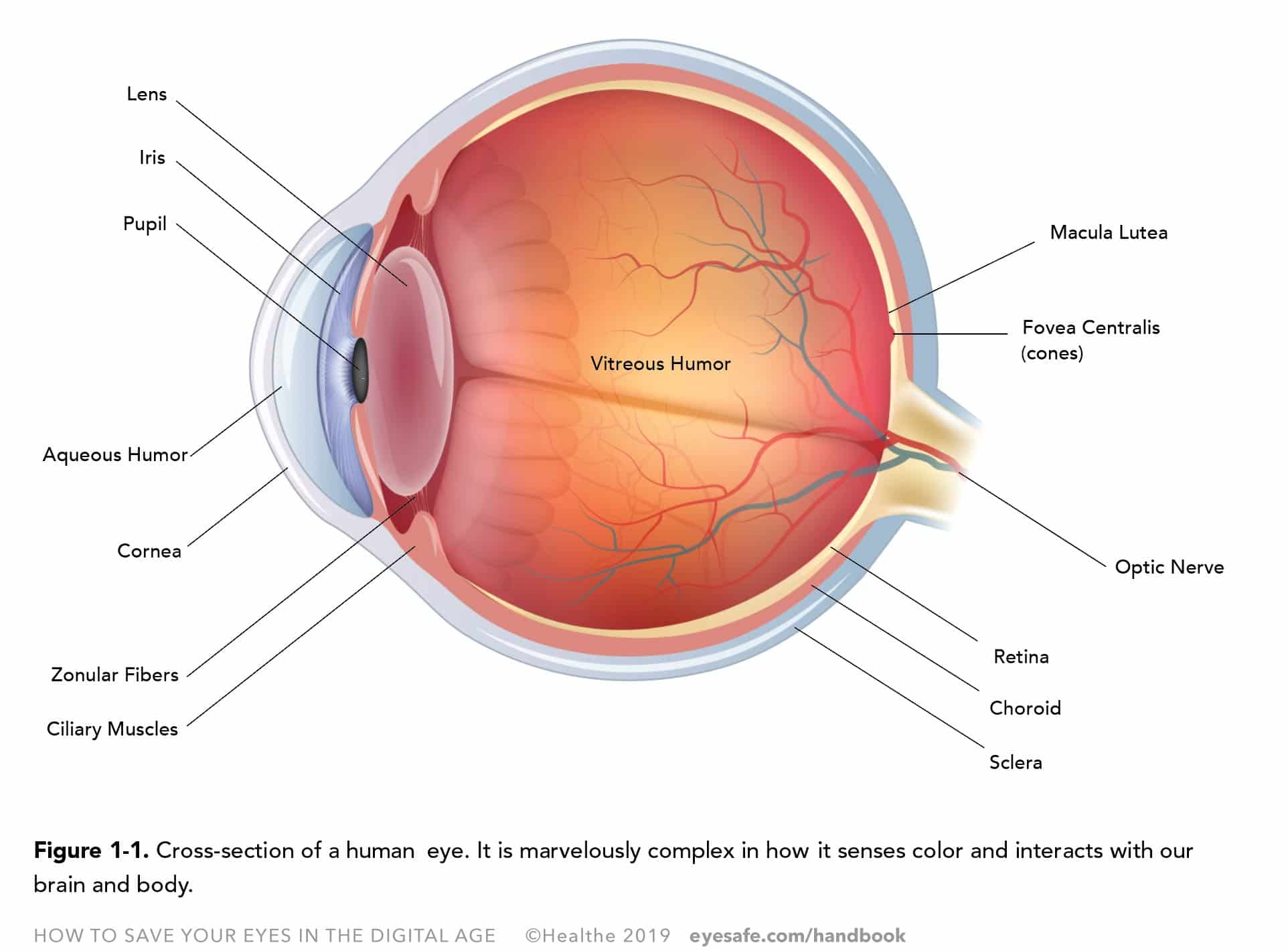
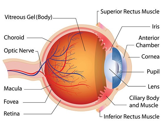
Where Is The Choroid Located In The Eye. Anatomically the outer portion of the eye is divided into three layers. Technically the choroid is the vascular part of the human eye that includes the connective tissue. Choroid the choroid proper located directly below the retina lines it from the outside. The vascular major blood vessel central layer of the eye lying between the retina and sclera.
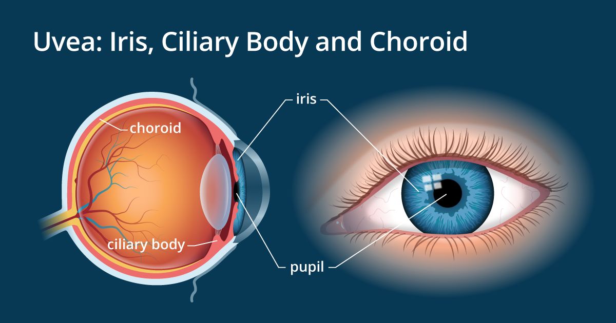 Iris And Uvea Of The Eye Allaboutvision Com From allaboutvision.com
Within this section of the eye there are four different layers. Its main purpose is to send oxygen and other nutrients to the retina. Eye develop ment starts at the end of the 4thweek when optical ditches appear in the cranial neural folds. The fibrous tunic cornea and sclera the vascular tunic choroid iris and ciliary body and the nervous tunic the eye is further divided into an anterior segment which contains the lens and structures anterior to it and a posterior segment which. The choroid is the vascular layer of the eye that lies between the retina and the sclera. Anatomically the outer portion of the eye is divided into three layers.
1 the choroid provides oxygen and nourishment to the outer layers of the retina.
Choroid the choroid proper located directly below the retina lines it from the outside. Technically the choroid is the vascular part of the human eye that includes the connective tissue. Eye develop ment starts at the end of the 4thweek when optical ditches appear in the cranial neural folds. Within this section of the eye there are four different layers. The eyes are paired sensory organs that enable vision. 1 the choroid provides oxygen and nourishment to the outer layers of the retina.
 Source: pinterest.com
The eyes are paired sensory organs that enable vision. The eyes are paired sensory organs that enable vision. Eye develop ment starts at the end of the 4thweek when optical ditches appear in the cranial neural folds. Its function is to provide nourishment to the outer layers of the retina through blood vessels. Technically the choroid is the vascular part of the human eye that includes the connective tissue.
 Source: youtube.com
Regulation of the flow of sunlight. 1 the choroid provides oxygen and nourishment to the outer layers of the retina. The choroid also known as the choroidea or choroid coat is the vascular layer of the eye containing connective tissues and lying between the retina and the sclera. Its main purpose is to send oxygen and other nutrients to the retina. Its function is to provide nourishment to the outer layers of the retina through blood vessels.
 Source: eyecancer.com
The fibrous tunic cornea and sclera the vascular tunic choroid iris and ciliary body and the nervous tunic the eye is further divided into an anterior segment which contains the lens and structures anterior to it and a posterior segment which. Choroid the choroid proper located directly below the retina lines it from the outside. The choroid also known as the choroid coat or choroidea is located between the retina and sclera. The choroid is the vascular layer of the eye that lies between the retina and the sclera. The important functions that are assigned to the choroid include.
 Source: fightingblindness.ie
Within this section of the eye there are four different layers. The choroid is thickest in the back of the eye where it is about 0 2 mm and narrows to 0 1 mm in the peripheral part of the eye. Anatomically the outer portion of the eye is divided into three layers. The choroid is a vascular layer that s to say it s made up of blood vessels and capillaries that lies between the white part of the eye the sclera and the internal retinal layer. Its function is to provide nourishment to the outer layers of the retina through blood vessels.
 Source: allaboutvision.com
The choroid is the vascular layer of the eye that lies between the retina and the sclera. Its function is to provide nourishment to the outer layers of the retina through blood vessels. Its main purpose is to send oxygen and other nutrients to the retina. The important functions that are assigned to the choroid include. The human choroid is thickest at the far extreme rear of the eye at 0 2 mm while in the outlying areas it narrows to 0 1 mm.
 Source: mayoclinic.org
The choroid is a vascular layer that s to say it s made up of blood vessels and capillaries that lies between the white part of the eye the sclera and the internal retinal layer. The eyes are paired sensory organs that enable vision. Technically the choroid is the vascular part of the human eye that includes the connective tissue. Development of the choroid the choroid is the middle layer of the eye is located in the posterior uveal. The choroid also known as the choroid coat or choroidea is located between the retina and sclera.
 Source: wisegeek.com
Choroid the choroid proper located directly below the retina lines it from the outside. The fibrous tunic cornea and sclera the vascular tunic choroid iris and ciliary body and the nervous tunic the eye is further divided into an anterior segment which contains the lens and structures anterior to it and a posterior segment which. Its function is to provide nourishment to the outer layers of the retina through blood vessels. The human choroid is thickest at the far extreme rear of the eye at 0 2 mm while in the outlying areas it narrows to 0 1 mm. After merging neural folds optical vesi cles are formed shaped as diverticula from forebrain wall.
 Source: cancer.gov
Regulation of the flow of sunlight. 1 it contains the retinal pigmented epithelial cells and provides oxygen and nourishment to the outer retina. The eyes are paired sensory organs that enable vision. The vascular major blood vessel central layer of the eye lying between the retina and sclera. Choroid the choroid proper located directly below the retina lines it from the outside.
 Source: eyecenternoco.com
Anatomically the outer portion of the eye is divided into three layers. The eyes are paired sensory organs that enable vision. 1 it contains the retinal pigmented epithelial cells and provides oxygen and nourishment to the outer retina. The fibrous tunic cornea and sclera the vascular tunic choroid iris and ciliary body and the nervous tunic the eye is further divided into an anterior segment which contains the lens and structures anterior to it and a posterior segment which. The choroid also known as the choroidea or choroid coat is the vascular layer of the eye containing connective tissues and lying between the retina and the sclera.
Source: aao.org
The fibrous tunic cornea and sclera the vascular tunic choroid iris and ciliary body and the nervous tunic the eye is further divided into an anterior segment which contains the lens and structures anterior to it and a posterior segment which. The human choroid is thickest at the far extreme rear of the eye at 0 2 mm while in the outlying areas it narrows to 0 1 mm. 1 it contains the retinal pigmented epithelial cells and provides oxygen and nourishment to the outer retina. Choroid the choroid proper located directly below the retina lines it from the outside. The fibrous tunic cornea and sclera the vascular tunic choroid iris and ciliary body and the nervous tunic the eye is further divided into an anterior segment which contains the lens and structures anterior to it and a posterior segment which.
 Source: en.wikipedia.org
The choroid is a vascular layer that s to say it s made up of blood vessels and capillaries that lies between the white part of the eye the sclera and the internal retinal layer. The choroid is thickest in the back of the eye where it is about 0 2 mm and narrows to 0 1 mm in the peripheral part of the eye. Technically the choroid is the vascular part of the human eye that includes the connective tissue. The vascular major blood vessel central layer of the eye lying between the retina and sclera. The choroid is a vascular layer that s to say it s made up of blood vessels and capillaries that lies between the white part of the eye the sclera and the internal retinal layer.
 Source: eyesafe.com
Within this section of the eye there are four different layers. The choroid also known as the choroidea or choroid coat is the vascular layer of the eye containing connective tissues and lying between the retina and the sclera. Within this section of the eye there are four different layers. After merging neural folds optical vesi cles are formed shaped as diverticula from forebrain wall. The choroid is a vascular layer that s to say it s made up of blood vessels and capillaries that lies between the white part of the eye the sclera and the internal retinal layer.
 Source: asrs.org
The choroid is a vascular layer that s to say it s made up of blood vessels and capillaries that lies between the white part of the eye the sclera and the internal retinal layer. Its function is to provide nourishment to the outer layers of the retina through blood vessels. Its main purpose is to send oxygen and other nutrients to the retina. Regulation of the flow of sunlight. Within this section of the eye there are four different layers.
 Source: researchgate.net
Anatomically the outer portion of the eye is divided into three layers. Development of the choroid the choroid is the middle layer of the eye is located in the posterior uveal. The choroid is a vascular layer that s to say it s made up of blood vessels and capillaries that lies between the white part of the eye the sclera and the internal retinal layer. The vascular major blood vessel central layer of the eye lying between the retina and sclera. Regulation of the flow of sunlight.
 Source: associatedretinaconsultants.com
1 it contains the retinal pigmented epithelial cells and provides oxygen and nourishment to the outer retina. The choroid is thickest in the back of the eye where it is about 0 2 mm and narrows to 0 1 mm in the peripheral part of the eye. 1 it contains the retinal pigmented epithelial cells and provides oxygen and nourishment to the outer retina. The vascular major blood vessel central layer of the eye lying between the retina and sclera. Anatomically the outer portion of the eye is divided into three layers.
If you find this site helpful, please support us by sharing this posts to your preference social media accounts like Facebook, Instagram and so on or you can also save this blog page with the title where is the choroid located in the eye by using Ctrl + D for devices a laptop with a Windows operating system or Command + D for laptops with an Apple operating system. If you use a smartphone, you can also use the drawer menu of the browser you are using. Whether it’s a Windows, Mac, iOS or Android operating system, you will still be able to bookmark this website.

